


1
Introduction
Bioelectromagnetism is a discipline that examines the electric, electromagnetic, and magnetic phenomena which arise in biological tissues. These phenomena include:
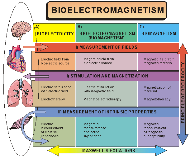
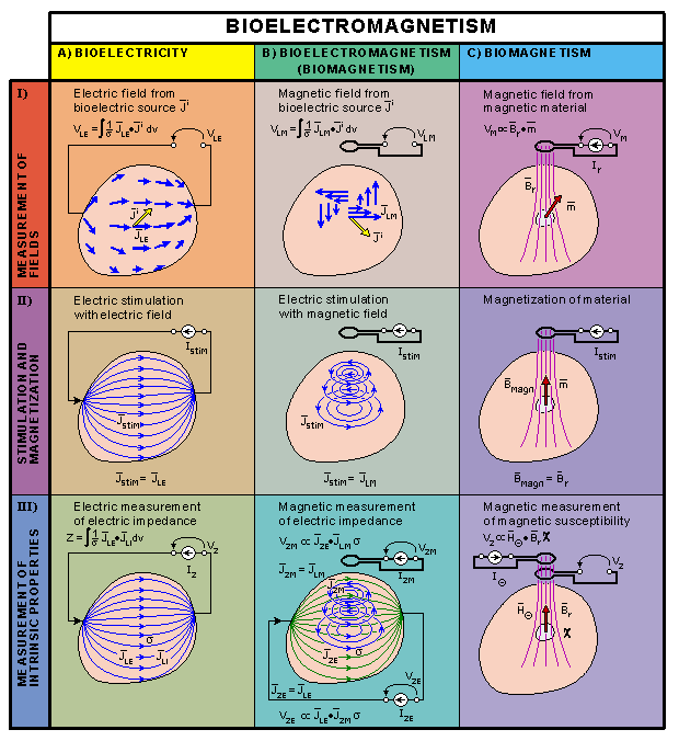
The discipline of bioelectromagnetism may be subdivided in many different ways. One such classification divides the field on theoretical grounds according to two universal principles: Maxwell's equations (the electromagnetic connection) and the principle of reciprocity. This philosophy is illustrated in Figure 1.2 and is discussed in greater detail below.
Fig. 1.3. Lead field theoretical approach to describe the subdivisions of bioelectromagnetism. The sensitivity distribution in the detection of bioelectric signals, the energy distribution in electric stimulation, and the distribution of measurement sensitivity of electric impedance are the same, owing to the principle of reciprocity. This is true also for the corresponding bioelectromagnetic and biomagnetic methods. Maxwell's equations tie time-varying electric and magnetic fields together so that when there are bioelectric fields there are also bioelectromagnetic fields, and vice versa.
Fig. 1.4. The first instrument to detect electricity was the electroscope invented by William Gilbert. (Gilbert 1600).
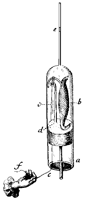
Fig. 1.5. Stimulation experiment of Jan Swammerdam in 1664. Touching the motoric nerve of a frog muscle (b) in a glass vessel (a) with silver wire (c) and a copper loop (d) produces stimulation of the nerve, which elicits a muscular contraction; however, it is uncertain as to whether the stimulation was produced as a result of electricity from the two dissimilar metals or from the mechanical pinching. See also text. (Swammerdam, 1738.).
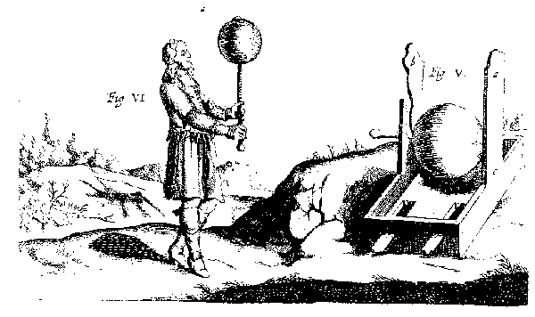
Fig. 1.6. Otto von Guericke constructed the first electric machine which included a sphere of sulphur with an iron axle. When rotating and rubbing the sphere it generated static electricity. (Guericke, 1672).
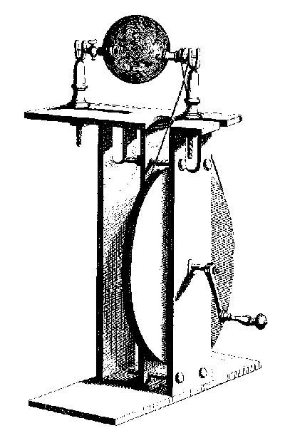
Fig. 1.7. Electric machine invented by Hauksbee in 1704. It had a sphere of glass rotated by a wheel. When the glass was rotated and rubbed it produced electricity continuously. If the glass was evacuated with air pump it generated brilliant light. (Hauksbee, 1709).
Fig. 1.8. The Leyden Jar, invented in 1745, was the first storage of electricity. It is formed by a glass bottle covered with metal foil on the inner and outer surfaces. (Krueger, 1746).
Fig. 1.9. Stimulation experiment of Luigi Galvani. The electrochemical behavior of two dissimilar metals [(zinc (Z) and copper (C)] in a bimetallic arch, in contact with the electrolytes of tissue, produces an electric stimulating current that elicits muscular contraction.
Fig. 1.10. Induction coil with hammer break. Electric current from the battery (E) is fed to the primary circuit of the induction coil (A). This current pulls the hammer with the magnetic field of the solenoid (close to G) and breaks the circuit with the contactor (D). Through the vibration of the hammer this breaking is continuous and it induces a high voltage alternating current in the secondary circuit in (A). This current is applied to the patient with electrodes (H)..
Fig. 1.12. Reconstruction of the first demonstration of the electromagnetic connection by Hans Christian Örsted in 1819. The battery generates an electric current I to flow in the circuit formed by a metal wire. This current induces a magnetic induction around the wire. The magnetic needle under the wire turns parallel to the direction of the magnetic induction demonstrating its existence. (Örsted, 1820a,b,c).
Fig. 1.13. (A) Astatic galvanometer invented by Nobili in 1825. He compensated for the effect of the Earth's magnetic field by placing two identical magnetic needles connected on the same suspension in opposite directions in the openings of a coil wound in the form of figure eight. (Nobili, 1825.)
(B) A technically more advanced version of the astatic galvanometer. Only one of the two identical (but opposite) needles is surrounded by a coil. The other needle serves as an indicator.
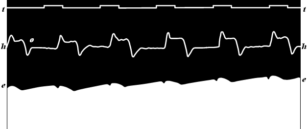
Fig. 1.16. The first recording of the human electrocardiogram by Augustus Waller (1887). The recording was made with a capillary electrometer. The ECG recording (e) is the borderline between the black and white areas. The other curve (h) is the apexcardiogram, a recording of the mechanical movement of the apex of the heart.
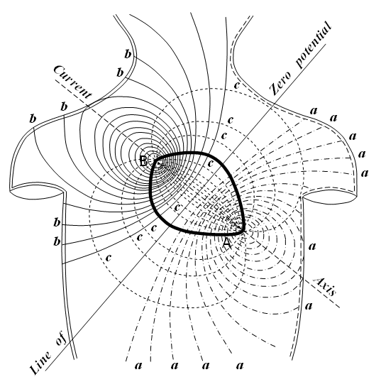
Fig. 1.17. Electric field of the heart on the surface of the thorax, recorded by Augustus Waller (1887). The curves (a) and (b) represent the recorded positive and negative isopotential lines, respectively. These indicate that the heart is a dipolar source having the positive and negative poles at (A) and (B), respectively. The curves (c) represent the assumed current flow lines..
Fig. 1.18. The monocardiogram by Mann. (Redrawn from Mann, 1920).
Table 1.6 Nobel prizes awarded in bioelectromagnetism and closely related subject areas
1.2 SUBDIVISIONS OF BIOELECTROMAGNETISM
1.2.1 Division on a Theoretical Basis
Maxwell's Equations
Maxwell's equations, i.e. the electromagnetic connection, connect time-varying electric and magnetic fields so that when there are bioelectric fields there always are also biomagnetic fields, and vice versa (Maxwell, 1865). Depending on whether we discuss electric, electromagnetic, or magnetic phenomena, bioelectromagnetism may be divided along one conceptual dimension (horizontally in Figure 1.2) into three subdivisions, namely
Reciprocity
Owing to the principle of reciprocity, the sensitivity distribution in the detection of bioelectric signals, the energy distribution in electric stimulation, and the sensitivity distribution of electric impedance measurements are the same. This is also true for the corresponding bioelectromagnetic and biomagnetic methods, respectively. Depending on whether we discuss the measurement of the field, of stimulation/magnetization, or the measurement of intrinsic properties of tissue, bioelectromagnetism may be divided within this framework (vertically in Figure 1.2) as follows:.
Description of the Subdivisions
The aforementioned taxonomy is illustrated in Figure 1.2 and a detailed description of its elements is given in this section.
Table 1.1 I ) Measurements of fields
(A) Bioelectricity (B) Bioelectromagnetism
(Biomagnetism)(C) Biomagnetism
Neural cells electroencephalography (EEG) magnetoencephalography (MEG) electroneurography (ENG) magnetoneurography (MNG) electroretinography (ERG) magnetoretinography (MRG) Muscle cells electrocardiography (ECG) magnetocardiography (MCG) electromyography (EMG) magnetomyography (MMG) Other tissue electro-oculography (EOG) magneto-oculography (MOG) electronystagmography (ENG) magnetonystagmography (MNG) magnetopneumogram magnetohepatogram
Table 1.2 II ) Stimulation and magnetization
(A) Bioelectricity (B) Bioelectromagnetism
(Biomagnetism)(C) Biomagnetism Stimulation patch clamp, voltage clamp electric stimulation of magnetic stimulation of electric cardiac pacing magnetic cardiac pacing electric cardiac defibrillation magnetic cardiac defibrillation Therapeutic applications electrotherapy electromagnetotherapy magnetotherapy electrosurgery Magnetization magnetization of
ferromagnetic material
Table 1.3 III ) Measurement of intrinsic properties
(A) Bioelectricity (B) Bioelectromagnetism
(Biomagnetism)(C) Biomagnetism electric measurement of
electric impedancemagnetic measurement of
electric impedancemeasurement of magnetic
susceptibilityimpedance cardiography magnetic susceptibility
plethysmographyimpedance pneumography magnetic remanence measurement impedance tomography impedance tomography magnetic resonance imaging (MRI) electrodermal response (EDR) Lead Field Theoretical Approach
As noted in the beginning of Section 1.2.1, Maxwell's equations connect time-varying electric and magnetic fields, so that when there are bioelectric fields there are also biomagnetic fields, and vice versa. This electromagnetic connection is the universal principle unifying the three subdivisions - A, B, and C - of bioelectromagnetism in the horizontal direction in Figure 1.2. As noted in the beginning of this section, the sensitivity distribution in the detection of bioelectric signals, the energy distribution in electric stimulation, and the sensitivity distribution of the electric impedance measurement are the same. All of this is true also for the corresponding bioelectromagnetic and biomagnetic methods, respectively. The universal principle that ties together the three subdivisions I, II, and III and unifies the discipline of bioelectromagnetism in the vertical direction in Figure 1.2 is the principle of reciprocity.
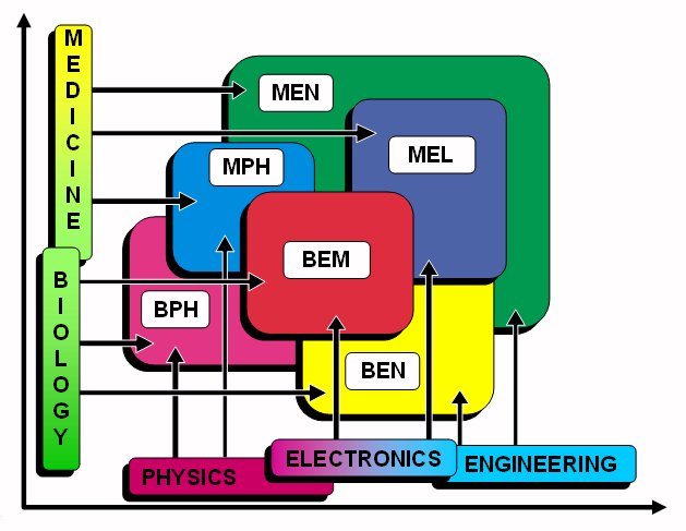
1.2.2 Division on an Anatomical Basis
Bioelectromagnetism can be classified also along anatomical lines. This division is appropriate especially when one is discussing clinical applications. In this case, bioelectromagnetism is subdivided according to the applicable tissue. For example, one might consider
1.2.3 Organization of this Book
Because it is inappropriate from a didactic perspective to use only one of the aforementioned divisional schemes (i.e.,division on a theoretical or an anatomical basis), both of them are utilized in this book. This book includes 28 chapters which are arranged into nine parts. Table 1.4 illustrates how these chapters fit into the scheme where by bioelectromagnetism is divided on a theoretical basis, as introduced in Figure 1.2.
Table 1.4 Organization of this book (by chapter number) according to the division of bioelectromagnetism on a theoretical basis.
(A) Bioelectricity
(B) Bioelectromagnetism
(Biomagnetism)(C) Biomagnetism
(I) Measurement of fields
Electric field from
bioelectric source
Magnetic field from
bioelectric source
Magnetic field from
magnetic material
04 Active behavior of the membrane
05 Physiology of the synapse and brain
06 Bioelectric behavior of the heart
07 Volume source and volume conductor
08 Source-field models
09 Bidomain model
11 Theoretical methods
13 Electroencephalography
15 12-lead ECG
16 Vectorcardiography
17 Other ECG systems
18 Distortion in ECG
19 ECG diagnosis
28 Electric signals of the eye
12 Theory of biomagnetic measurements
14 Magnetoencephalography
20 Magnetocardiography
Not discussed
(II) Stimulation and magnetization
Electric stimulation
with electric field
Electric stimulation
with magnetic field
Magnetization of
material
03 Subthreshold membrane phenomena
21 Functional electric stimulation
23 Cardiac pacing
24 Cardiac defibrillation
22 Magnetic stimulation
Not discussed
(III) Measurement of intrinsic properties
Electric measurement of
electric impedanceMagnetic measurement of
electric impedanceMagnetic measurement of
magnetic susceptibility
25 Impedance plethysmography
26 Impedance tomography
27 Electrodermal response
26 Magnetic measurement of
electric impedance tomography
Not discussed
1.3 IMPORTANCE OF BIOELECTROMAGNETISM
Why should we consider the study of electric and magnetic phenomena in living tissues as a separate discipline? The main reason is that bioelectric phenomena of the cell membrane are vital functions of the living organism. The cell uses the membrane potential in several ways. With rapid opening of the channels for sodium ions, the membrane potential is altered radically within a thousandth of a second. Cells in the nervous system communicate with one another by means of such electric signals that rapidly travel along the nerve processes. In fact, life itself begins with a change in membrane potential. As the sperm merges with the egg cell at the instant of fertilization, ion channels in the egg are activated. The resultant change in the membrane potential prevents access of other sperm cells.
1.4 SHORT HISTORY OF BIOELECTROMAGNETISM
1.4.1 The First Written Documents and First Experiments in Bioelectromagnetism
The first written document on bioelectric events is in an ancient Egyptian hieroglyph of 4000 B.C. The hieroglyph describes the electric sheatfish (catfish) as a fish that "releases the troops." Evidently, when the catch included such a fish, the fish generated electric shocks with an amplitude of more than 450 V, which forced the fishermen to release all of the fish. The sheatfish is also illustrated in an Egyptian sepulcher fresco (Morgan, 1868).
1.4.2 Electric and Magnetic Stimulation
Systematic application of electromedical equipment for therapeutic use started in the 1700s. One can identify four different historical periods of electromagnetic stimulation, each based on a specific type or origin of electricity. These periods are named after Benjamin Franklin (American; 1706-1790), Luigi Galvani (Italian; 1737-1798), Michael Faraday (British; 1791-1867), and Jacques Arsčne d'Arsonval (French; 1851-1940), as explained in Table 1.5. These men were the discoverers or promoters of different kinds of electricity: static electricity, direct current, induction coil shocks, and radiofrequency current, respectively (Geddes, 1984a).
Table 1.5. Different historical eras of electric and
electromagnetic stimulation.
Scientist Lifetime Historical era Benjamin Franklin 1706-1790 static electricity Luigi Galvani 1737-1798 direct current Michael Faraday 1791-1867 induction coil shocks Jacques d'Arsonval 1851-1940 radiofrequency current 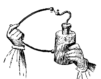
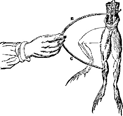
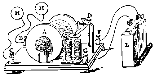
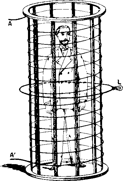
Fig. 1.11. d'Arsonval's great solenoid. (d'Arsonval, 1893).
1.4.3 Detection of Bioelectric Activity
The connection between electricity and magnetism was discovered in 1819 by Hans Christian Örsted (Danish; 1777-1851). Örsted conducted his first experiment during his lecture at the University of Copenhagen. Passing an electric current through a wire above a magnetic needle, he forced the needle to move to the direction normal to the wire (see Figure 1.12) (Örsted, 1820a,b,c). By reversing the direction of the electric current, he reversed the direction of the needle deflection. (The magnetic needle, i.e. the compass, was invented in China about A.D. 100 and is the first detector of magnetic field.)
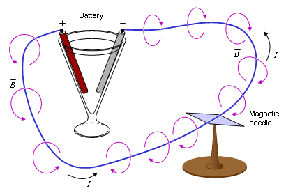
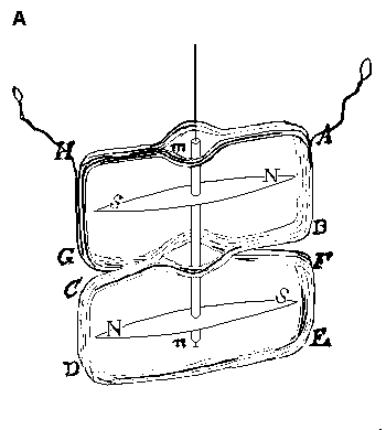
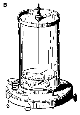
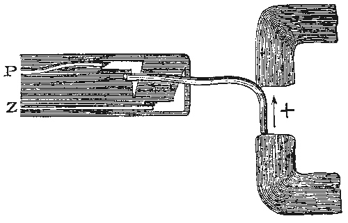
Fig. 1.14. Du Bois-Reymond's apparatus for studying effect of continuous current on nerve.
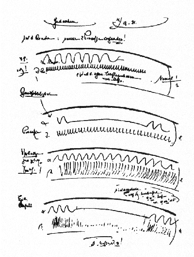
Fig. 1.15. A page from Berger's notebook illustrating early recordings of the human EEG.
1.4.4 Modern Electrophysiological Studies of Neural Cells
The term neuron was first applied to the neural cell in 1891 by Heinrich Wilhelm Gottfried Waldeyer (German; 1837-1921). Basic research into the study of neurons was undertaken at the end of the nineteenth century by August Forel (Swiss; 1848-1931), Wilhelm His, Sr. (Swiss; 1831-1904), and Santiago Ramón y Cajal (Spanish; 1852-1934). According to their theory, it is the neural cell that is the functional unit in the nervous system. (In 1871, Santiago Ramón y Cajal also discovered that neurons could be selectively stained with a special silver preparation.)
1.4.5 Bioelectromagnetism
As mentioned in Section 1.4.3, the connection between electricity and magnetism was experimentally discovered in 1819 by Hans Christian Örsted. French scientists Jean Baptiste Biot (1774- 1862) and Félix Savart (1791-1841) proved that the force between a current-carrying helical wire and a magnet pole is inversely proportional to the distance between them (Biot, 1820). André Marie Ampčre (French; 1775-1836) showed that a current-carrying helical wire, which he called the solenoid, behaved magnetically as a permanent magnet (Ampčre, 1820), hence linking the electric current to the production of a magnetic field. Ampčre also developed the mathematical theory of electrodynamics (Ampčre, 1827). The electromagnetic connection was theoretically formulated in 1864 by James Clerk Maxwell (British; 1831-79), who developed equations that link time-varying electricity and magnetism (Maxwell, 1865). Since Örsted's discovery, electromagnetic interdependence has been widely utilized in a large variety of devices. Examples of these include those used for the measurement of electric current (galvanometers and ammeters), electric generators, electric motors, and various radiofrequency devices. However, biomagnetic signals were not detected for a long time because of their extremely low amplitude.
Fig. 1.19. Detection of the first biomagnetic signal, the magnetocardiogram (MCG), by Baule and McFee. (Redrawn from Baule and McFee, 1963.).
1.4.6 Theoretical Contributions to Bioelectromagnetism
The German scientist and philosopher Hermann Ludwig Ferdinand von Helmholtz (1821-1894) made the earliest significant contributions of the theory of bioelectromagnetism. A physician by education and, in 1849, appointed professor of physiology at Königsberg, he moved to the chair of physiology at Bonn in 1855. In 1871 he was awarded the chair of physics at the University of Berlin, and in 1888 was also appointed the first director of Physikalisch-Technische Bundesanstalt in Berlin.
1. The demonstration that axons are processes of nerve cell bodies (1842)
2. The establishment of the law of conservation of energy (the First Law of Thermodynamics) (1847)
3. The invention of the myograph and the first measurement of the conduction velocity of a motor nerve axon (1850)
4. The concept of double layer source (1853)
5. The solid angle theorem for electric potentials
6. The principle of superposition (1853)
7. The reciprocity theorem (1853)
8. The insolvability of the inverse problem (1853)
9. Helmholtz's theorem concerning the independence of flow and vortex sources
10. The Helmholtz coils (applied in biomagnetic instrumentation)
1.4.7 Summary of the History of Bioelectromagnetism
The history of bioelectromagnetism is summarized chronologically in Figure 1.20. The historical events are divided into four groups: theory, instrumentation, stimulation, and measurements. This figure should serve as a useful overview for our readers and help them recognize how one contribution follows from an earlier one and how the development of an entire discipline thereby takes place. From this figure we may summarize the following thoughts.
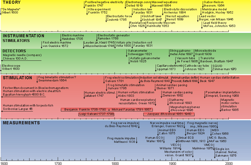
Fig. 1.20. Chronology of the history of bioelectromagnetism. The historical events are divided into four groups: theory, instrumentation, stimulation, and measurements..
1.5 NOBEL PRIZES IN BIOELECTROMAGNETISM
The discipline of bioelectromagnetism is strongly reflected in the work of many Nobel laureates. It should be noted that 16 Nobel prizes have been given for contributions to the discipline of bioelectromagnetism and closely related subjects. Of these prizes, 12 were in physiology or medicine; four were in chemistry. Although some perhaps do not directly concern bioelectromagnetism, they are very closely related. Since several individuals may have shared an award, the actual number of Nobel laureates is 28. The large number of these Nobel laureates shows that bioelectromagnetism is recognized as a very important discipline. Nobel laureates associated with bioelectromagnetism are listed in Table 1.6.
*) Nobel Prize in chemistry. All other prizes were received in physiology or medicine.
Year Name of recipient Nationality Subject of research 1901 Jacobus van't Hoff *) The Netherlands laws of chemical dynamics
and osmotic pressure1903 Svante Arrhenius *) Sweden theory of electrolytic
dissociation1906 Camillo Golgi
Santiago Ramón y Cajal Italy
Spainwork on the structure of
nervous system1920 Walther Nernst *) Germany work in thermochemistry 1924 Willem Einthoven The Netherlands discovery of electro-
cardiogram mechanism1932 Edgar Douglas Adrian
Sir Charles SherringtonBritain
Britaindiscoveries regarding
function of neurons1936 Sir Henry Hallet Dale
Otto LoewiBritain
Germanywork on chemical trans-
mission of nerve impulses1944 Joseph Erlanger
Herbert Spencer GasserU.S.
U.S.researches on differentiated
functions of nerve fibers1949 Walter Rudolf Hess Switzerland discovery of function of
middle brain1961 Georg von Békésy U.S. discoveries of the physical
mechanism of the inner ear1963 Sir John Eccles
Alan Lloyd Hodgkin
Andrew Fielding HuxleyAustralia
Britain
Britainstudy of the transmission
of nerve impulses along a
nerve fibre1967 Ragnar Arthur Granit
Haldan Keffer Hartline
George WaldFinland
U.S.
U.S.discoveries about chemical
and physiological visual
processes in the eye1968 Lars Onsager *) U.S. work on theory of thermo-
dynamics of irreversible
processes1970 Julius Axelrod
Sir Bernard Katz
Ulf von EulerU.S.
Britain
Swedendiscoveries concerning the
chemistry of nerve
transmission1981 David Hunter Hubel
Torsten Nils WieselU.S.
Swedendiscoveries concerning
information processing
in the visual system1991 Erwin Neher
Bert SakmannGermany
Germanydiscoveries concerning
the function of single
ion channels in cells1997 Paul D. Boyer
John E. Walker
Jens C. Skou *)U.S.
U.K.
Denmarkthe enzymatic mechanism
underlying the synthesis of ATP;
discovery of an ion-transporting
enzyme, Na+, K+ -ATPase2003 Peter Agre
Roderick MacKinnon *)U.S.
U.S.discoveries concerning
channels in cell membranes
Ader C (1897): Sur un nouvel appareil enregistreur pour cables sousmarins. Compt. rend. Acad. Sci. (Paris) 124: 1440-2.
Adrian ED (1914): The all-or-none principle in nerve. J. Physiol. (Lond.) 47: 460-74.
Adrian ED, Lucas K (1912): On the summation of propagated disturbances in nerve and muscle. J. Physiol. (Lond.) 44: 68-124.
Aldini G (1804): Essai Théorique et Expérimental sur le Galvanisme, Vol. 2, Fournier, Paris.
Ampčre AM (1820): Du mémoire sur l'action mutuelle entre deux courans électriques, entre un courant électrique et un aimant ou le globe terrestre, et entre deux aimans. Ann. Chim. Phys. 15: 170-218.
Ampčre AM (1827): Mémoire sur la théorie mathématique des phénomčnes électro-dynamiques uniquemant déduit de l'éxperience. Mém. de l'Institut 6: 175-386.
d'Arsonval JA (1893): Action physiologique de courants alternatifs a grande frequence. Arch. Physiol. Norm. et Pathol. 5: 401-8, 780-90.
d'Arsonval JA (1896): Dispositifs pour la mésure des courants alternatifs de toutes fréquences. C. R. Soc. Biol. (Paris) 2: 450-1.
Barker AT, Jalinous R, Freeston IL (1985): Non-invasive magnetic stimulation of human motor cortex. Lancet 1:(8437) 1106-7.
Bartholow R (1881): Electro-Therapeutics, Philadelphia.
Baule GM, McFee R (1963): Detection of the magnetic field of the heart. Am. Heart J. 55:(7) 95-6.
Beck A (1888): O pobudiwosci róznych miejsc tego samego nerwu. Rozpr. Wydz. mat.-przyr. polsk. Akad. Um. 15: 165-95. (On the excitability of the various parts of the same nerve).
Beck A (1891): The determination of localization in the brain and spinal cord by means of electrical phenomena. Polska Akademija Umiejetnosci, Series 2, pp. 187-232. (thesis)
Beck CS, Pritchard WH, Feil HS (1947): Ventricular fibrillation of long duration abolished by electric shock. JAMA, J. Am. Med. Assoc. 135: 985.
Berger H (1929): Über das Elektroenkephalogram des Menschen. Arch. f. Psychiat. 87: 527-70.
Biot JB (1820): Note sur le magnétisme de la pile de Volta. Ann. Chim. Phys. 15: 222-4.
du Bois-Reymond EH (1843): Vorläufiger Abriss einer Untersuchung ueber den sogenannten Froschstrom und ueber die elektromotorischen Fische. Ann. Physik und Chemie 58: 1-30.
du Bois-Reymond EH (1848): Untersuchungen ueber thierische Elektricität, Vol. 1, 56+743 pp. G Reimer, Berlin.
du Bois-Reymond EH (1849): Untersuchungen ueber thierische Elektricität, Vol. 2, pp. 393-4. G. Reimer, Berlin.
Brazier MAB (1959): The historical development of neurophysiology. In Handbook of Physiology. Section I: Neurophysiology, Vol. I, ed. IJ Field, HW Magoun, VE Hall, pp. 1-58, American Physiological Society, Washington.
Burger HC, van Milaan JB (1946): Heart vector and leads - I. Br. Heart J. 8:(3) 157-61.
Caton R (1875): The electric currents of the brain. Br. Med. J. 2: 278.
Cohen D (1968): Magnetoencephalography, evidence of magnetic fields produced by alpha-rhythm currents. Science 161: 784-6.
Cohen D (1972): Magnetoencephalography: Detection of brain's electric activity with a superconducting magnetometer. Science 175:(4022) 664-6.
Cohen D, Edelsack EA, Zimmerman JE (1970): Magnetocardiograms taken inside a shielded room with a superconducting point-contact magnetometer. Appl. Phys. Letters 16: 178-280.
Einthoven W (1908): Weiteres über das Elektrokardiogram. Pflüger Arch. ges. Physiol. 122: 517-48.
Einthoven W, Fahr G, de Waart A (1913): Über die Richtung und die Manifeste Grösse der Potentialschwankungen im mennschlichen Herzen und über den Einfluss der Herzlage auf die form des Elektrokardiogramms. Pflüger Arch. ges. Physiol. 150: 275-315.
Faraday M (1834): Experimental researches on electricity, 7th series. Phil. Trans. R. Soc. (Lond.) 124: 77-122.
Furman S, Schwedel JB (1959): An intracardiac pacemaker for Stokes-Adams seizures. N. Engl. J. Med. 261:(5 Nov) 943-8.
Gabor D, Nelson CV (1954): Determination of the resultant dipole of the heart from measurements on the body surface. J. Appl. Phys. 25:(4) 413-6.
Galvani L (1791): De viribus electricitatis in motu musculari. Commentarius. De Bononiesi Scientarium et Ertium Instituto atque Academia Commentarii 7: 363-418. (Commentary on the effects of electricity on muscular motion. Burndy Library edition, 1953, Norwalk, Conn.).
Gasser HS, Erlanger J (1922): A study of the action currents of the nerve with the cathode ray oscillograph. Am. J. Physiol. 62: 496-524.
Geddes LA (1976): Kouwenhoven WB. Med. Instrum. 10:(2) 141-3.
Gilbert W (1600): De Magnete, Magneticisque Corporibus, et de Magno Magnete Tellure; Physiologica Nova Plumiris et Argumentis et Experimentis Demonstrata, Peter Short, London. (Transl. SP Thompson, London: The Gilbert Club, 1900: facsimile ed. New York: Basic Books, 1958: transl. PF Mottelay, 1893, facsimile ed.: Dover, New York, 1958.)
Granit R (1955): Receptors and Sensory Perception, 369 pp. Yale University Press, New Haven.
Green T (1872): On death from chloroform: Its prevention by a galvanism. Br. Med. J. 1:(May 25) 551-3.
von Guericke O (1672): Experimenta Nova (Ut Vocantur) Magdeburgica, Amsterdam.
Hauksbee F (1709): Physico-Mechanical Experiments, 1st ed., London.
Helmholtz HLF (1842): De fabrica systematis nervosi evertebratorum. Berlin, Thesis, (Structure of the nervous system in invertebrates.) (Dr. Phil. thesis)
Helmholtz HLF (1847): Über die Erlangung der Kraft, Berlin. (On the conservation of energy)
Helmholtz HLF (1850): Über die Fortpflanzungsgeschwindigkeit der Nervenreizung. Arch. anat. physiol. wiss. Med. : 71-3. (On the speed of propagation of nerve stimulation).
Helmholtz HLF (1853): Ueber einige Gesetze der Vertheilung elektrischer Ströme in körperlichen Leitern mit Anwendung auf die thierisch-elektrischen Versuche. Ann. Physik und Chemie 89: 211-33, 354-77.
Helmholtz HLF (1863): Die Lehre von den Tonempfindungen als Physiologische Grundlage für die Theorie der Musik, Braunschweig. (On the sensations of tone)
Hodgkin AL, Huxley AF (1952): A quantitative description of membrane current and its application to conduction and excitation in nerve. J. Physiol. (Lond.) 117: 500-44.
Hyman S (1932): Resuscitation in stopped heart. Arch. Int. Med. 50: 283-305.
Jallabert J (1748): Expériences Sur L'électricité Avec Quelques Conjectures Sur La Cause De Ses Effets, Geneva.
Johnson JB (1921): A low voltage cathode ray oscillograph. Physical Rev. 17: 420-1.
Kellaway P (1946): Bull. Hist. Med. 20: 112-37.
Krueger J (1746): Beschichte der Erde, Lubetvatbischen Buchhandlung, Helmstädt.
Licht S (1967): Therapeutic Electricity and Ultraviolet Radiation, Baltimore.
Lippman GJ (1873): Beziehungen zwischen der Capillaren und elektrischen Erscheinungen. Ann. Physik und Chemie (Series 2) 149: 546-61.
Maniewski R, Katila T, Poutanen T, Siltanen P, Varpula T, Wikswo JP (1988): Magnetic measurement of cardiac mechanical activity. IEEE Trans. Biomed. Eng. 35:(9) 662-70.
Mann H (1920): A method for analyzing the electrocardiogram. Arch. Int. Med. 25: 283-94.
Marey EJ, Lippman GJ (1876): Inscription photographique des indications de l'électromčtre de Lippman. Compt. rend. Acad. Sci. (Paris) 83: 278-80.
Matteucci C (1838): Sur le courant électrique oů propre de la grenouille. Second memoire sur l'électricité animale, faisant suite ŕ celui sur la torpille. Ann. Chim. Phys. (2čme serie), 67: 93-106.
Matteucci C (1842): Deuxičme mémoire sur le courant électrique propre de la grenouille et sur celui des animaux ŕ sang chaud (1). Ann. Chim. Phys. (3čme serie,) 6: 301-39.
Maxwell J (1865): A dynamical theory of the electromagnetic field. Phil. Trans. R. Soc. (Lond.) 155: 459-512.
McFee R, Johnston FD (1953): Electrocardiographic leads I. Introduction. Circulation 8:(10) 554-68.
McFee R, Johnston FD (1954a): Electrocardiographic leads II. Analysis. Circulation 9:(2) 255-66.
McFee R, Johnston FD (1954b): Electrocardiographic leads III. Synthesis. Circulation 9:(6) 868-80.
Morgan CE (1868): Electro-Physiology and Therapeutics, Williams, Wood, New York.
Neher E, Sakmann B (1976): Single-channel currents recorded from membrane of denervated frog muscle fibers. Nature 260: 799-802.
Nobili L (1825): Ueber einen neuen Galvanometer. J. Chem. und Physik 45: 249-54.
Polson MJ, Barker AT, Freeston IL (1982): Stimulation of nerve trunks with time-varying magnetic fields. Med. & Biol. Eng. & Comput. 20:(2) 243-4.
Prevost JL, Battelli F (1899): On some effects of electric discharges on the heart of mammals. Compt. rend. Acad. Sci. (Paris) 129: 943-8.
de la Rive A (1853): A Treatise on Electricity, London.
Schweigger JSC (1821): Elektromagnetischer Multiplikator. J. Chem. und Physik 31: 35-41.
Sherrington CS (1897): The central nervous system. In A Textbook of Physiology, Vol. 3, ed. M Forster, MacMillan, London.
Smith JA, (Transl.) (1931): De anima. In The Works of Aristotle, Vol. 3, ed. WD Ross, p. 405, Oxford University Press, Oxford.
Steiner F (1871): Über die Elektropunctur des Herzens als Wiederbelebungsmittel in der Chloroformsyncope. Archiv. f. klin. Chir. 12: 748-80.
Swammerdam J (1738a): Biblia Naturae, Vol. 2, ed. H. Boerhaave, Leyden.
Swammerdam J (1738b): Biblia Naturae, Vol. 2, ed. H. Boerhaave, pp. 839-50. Leyden.
Volta A (1800): On the electricity excited by the mere contact of conducting substances of different kinds. Phil. Trans. R. Soc. (Lond.) 90: 403-31. (In French.).
Waller AD (1887): A demonstration on man of electromotive changes accompanying the heart's beat. J. Physiol. (Lond.) 8: 229-34.
Wikswo JP, Barach JP, Freeman JA (1980): Magnetic field of a nerve impulse: First measurements. Science 208: 53-5.
Williams HB (1914): On the cause of the phase difference frequently observed between homonymous peaks of the electrocardiogram. Am. J. Physiol. 35: 292-300.
York DH (1987): Review of descending motor pathways involved with transcranial stimulation. Neurosurg. 20:(1) 70-3.
Ziemssen H (1882): Studien über die Bewegungsvorgange am menschlichen Herzen. Deuts. Archiv f. klin. Med. 30: 270-303.
Zimmerman JE, Thiene P, Hardings J (1970): Design and operation of stable r-f biased superconducting point-contact quantum devices. J. Appl. Phys. 41: 1572.
Zoll PM (1952): Excitation of the heart in ventricular standstill by external electric stimulation. N. Engl. J. Med. 247: 768-71.
Örsted HC (1820a): Experimenta circa effectum conflictus electrici in acum magneticam. J. F. Chem. Phys. 29: 275-81.
Örsted HC (1820b): Galvanic magnetsim. Phil. Mag. 56: 394.
Örsted HC (1820c): Neuere elektro-magnetische Versuche. J. Chem. und Physik 29: 364-9.
Brazier MAB (1959): The historical development of neurophysiology. In Handbook of Physiology. Section I: Neurophysiology, Vol. I, ed. IJ Field, HW Magoun, VE Hall, pp. 1-58, American Physiological Society, Washington.
Brazier MA (1988): A History of Neurophysiology in the 19th Century, 265 pp. Raven Press, New York.
Gandhi OP (ed.) (1990): Biological Effects and Medical Applications of Electromagnetic Energy, (Series ed, A Nordgraaf: Biophysics and Bioengineering Series.) 573 pp. Prentice Hall, Englewood Cliffs, N.J.
Geddes LA (1984a): The beginnings of electromedicine. IEEE Eng. Med. Biol. Mag. 3:(4) 8-23.
Geddes LA (1984b): A short history of the electrical stimulation of excitable tissue: Including electrotherapeutic applications. Physiologist 27(Suppl.):(1) 15-265.
McNeal DR (1977): 2000 years of electrical stimulation. In Functional Electrical Stimulation. Applications in Neural Prostheses. Biomedical Engineering and Instrumentation Series, Vol. 3, ed. FT Hambrecht, JB Reswick, pp. 3-35, Marcel Dekker, New York and Basel.
Mottelay PF (1975): Bibliographical History of Electricity and Magnetism, 673 pp. Charles Griffin, New York.
Rautaharju PM (1987): A hundred years of progress in electrocardiography, 1: Early contributions from Waller to Wilson. Can. J. Cardiol. 3:(8) 362-74.
Rautaharju PM (1988): A hundred years of progress in electrocardiography, 2: The rise and decline of vectorcardiography. Can. J. Cardiol. 4:(2) 60-71.
Reilly JP (1992): Electrical Stimulation & Electropathology, 504 pp. Cambridge University Press, Cambridge.
Rowbottom M, Susskind C (1984): Electricity and Medicine. History of Their Interaction, 303 pp. San Francisco Press, San Francisco.
Wasson T (ed.) (1987): Nobel Prize Winners, 1165 pp. H. W. Wilson, New York.