11
Theoretical Methods for
Analyzing Volume Sources
and Volume Conductors
The first two theoretical methods of this chapter (solid angle theorem and Miller-Geselowitz model) are used to evaluate the electric field in a volume conductor produced by the source - that is, to solve the forward problem. After this discussion is a presentation of methods used to evaluate the source of the electric field from measurements made outside the source, inside or on the surface of the volume conductor - that is, to solve the inverse problem. These methods are important in designing electrode configurations that optimize the capacity to obtain the desired information.
The solid angle theorem was developed by the German physicist Hermann von Helmholtz in the middle of the nineteenth century. In this theory, a double layer is used as the source. Although this topic was introduced in Chapter 8, we now examine the structure of a double layer in somewhat greater detail.
Of course, the double layer arises only in the limit that d
 In fact, application of each of the following methods usually results in a particular ECG-lead system. These lead systems are not discussed here in detail because the purpose of this chapter is to show that these methods of analysis form an independent theory of bioelectricity that is not limited to particular ECG applications.
The biomagnetic fields resulting from the electric activity of volume sources are discussed in detail in Chapter 12.
In fact, application of each of the following methods usually results in a particular ECG-lead system. These lead systems are not discussed here in detail because the purpose of this chapter is to show that these methods of analysis form an independent theory of bioelectricity that is not limited to particular ECG applications.
The biomagnetic fields resulting from the electric activity of volume sources are discussed in detail in Chapter 12.
11.2 SOLID ANGLE THEOREM
11.2.1 Inhomogeneous Double Layer
PRECONDITIONS:
SOURCE: Inhomogeneous double layer
CONDUCTOR: Infinite, homogeneous, (finite, inhomogeneous)
Suppose that a point current source and a current sink (i.e., a negative source) of the same magnitude are located close to each other. If their strength is i and the distance between them is d, they form a dipole moment id as discussed in Section 8.2.2. Consider now a smooth surface of arbitrary shape lying within a volume conductor. We can uniformly distribute many such dipoles over its surface, with each dipole placed normal to the surface. In addition, we choose the dipole density to be a well-behaved function of position - that is, we assume that the number of dipoles in a small area is great enough so that the density of dipoles can be well approximated with a continuous function. Such a source is called a double layer (Figure 11.1). If it is denoted by p(S) , then p(S) denotes a dipole moment density (dipole moment per unit area) as a function of position, while its direction is denoted by , the surface normal. With this notation, p(S)d
 is a dipole whose magnitude is p(S)dS, and its direction is normal to the surface at dS.
An alternative point of view is to recognize that on one side of the double layer, the sources form a current density J [A/m2] whereas on the other side the sinks form a current density -J [A/m2], and that the conducting sheet between the surfaces of the double layer has a resistivity r. The resistance across this sheet (of thickness d) for a unit cross-sectional area is
is a dipole whose magnitude is p(S)dS, and its direction is normal to the surface at dS.
An alternative point of view is to recognize that on one side of the double layer, the sources form a current density J [A/m2] whereas on the other side the sinks form a current density -J [A/m2], and that the conducting sheet between the surfaces of the double layer has a resistivity r. The resistance across this sheet (of thickness d) for a unit cross-sectional area is
 R = rd
(11.1)
where R = double layer resistance times unit area [Wm˛] r = resistivity of the medium [Wm] d = thickness of the double layer [m]  0 while J
  such that Jd p remains finite.
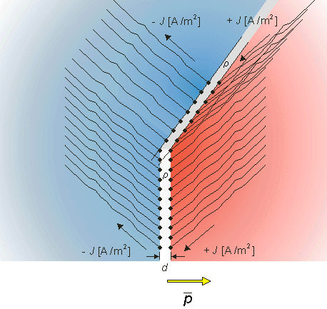
Fig. 11.1. Structure of a double layer. The double layer is formed when the dipole density increases to the point that it may be considered a continuum. In addition, we require that J
, d 0, and Jd p.From Ohm's law we note that the double layer has a potential difference of
| Vd = F1 - F2 = Jrd | (11.2) |
| where | Vd | = voltage difference over the double layer [V] |
| F1, F2, | = potentials on both sides of the double layer [V] | |
| J | = double layer current density [A/m2] | |
| r | = resistivity of the medium [Wm] | |
| d | = double layer thickness [m] |
By definition, the double layer forms a dipole moment per unit surface area of
| p = Jd | (11.3) |
| where | p | = dipole moment per unit area [A/m] |
| J | = double layer current density [A/m2] | |
| d | = double layer thickness [m] |
As noted, in the general case (nonuniform double layer), p and J are functions of position. Strictly we require d
since the direction of
Thus
0 while J such that Jd = p remains finite. (In the case where d is not uniform, then for Equation 11.2 to be a good approximation it is required that DF not vary significantly over lateral distances several times d.)
Since 
 p is the dipole moment per unit area (with the direction from negative to positive source), dS is an elementary dipole. Its field, given by Equation 8.12 is:
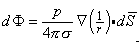
(11.4) and d are the same. Now the solid angle dW, as defined by Stratton (1941), is:
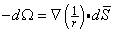
(11.5)
(11.6)
The Polarity of the Potential Field
We discuss shortly the polarity of the potential field generated by a double layer. This will clarify the minus sign in Equations 11.5 and 11.6.
A uniform double layer exhibits some interesting properties that are discussed here in this section.
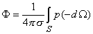
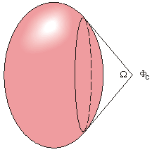
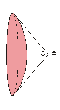
The double layer generates a potential field given by Equation 11.6, where dW is the element of solid angle, as seen from the field point as the point of observation (Figure 11.2). This figure provides an interpretation of the solid angle as a measure of the opening between rays from the field point to the periphery of the double layer, a form of three-dimensional angle. Equation 11.6 has a particularly simple form, which readily permits an estimation of the field configuration arising from a given double layer source function.
This result was first obtained by Helmholtz, who showed that it holds for an infinite, homogeneous, isotropic, and linear volume conductor. Later the solid angle theorem was also applied to inhomogeneous volume conductors by utilizing the concept of secondary sources. As discussed in Section 7.2.3, the inhomogeneous volume conductor may be represented as a homogeneous volume conductor including secondary sources at the sites of the boundaries. Now the potential field of a double layer source in an inhomogeneous volume conductor may be calculated with the solid angle theorem by applying it to the primary and secondary sources in a homogeneous volume conductor.
If the double layer is uniform, then the field point's potential is proportional to the total solid angle subtended at the field point. It is therefore of interest to be able to determine this solid angle. One useful approach is the following: From the field point, draw lines (rays) to the periphery of the double layer surface. Now construct a unit sphere centered at the field point. The area of the sphere surface intercepted by the rays is the solid angle. If the negative sources associated with the double layer face the field point, then the solid angle will be positive, according to Equation 11.5. This polarity arises from the purely arbitrary way in which the sign in Equation 11.5 was chosen. Unfortunately, the literature contains both sign choices in the definition of the solid angle (in this book we adopted the one defined by Stratton, 1941).
For example, suppose a uniform double layer is a circular disk centered at the origin, whose dipoles are oriented in the x direction. For a field point along the positive x-axis, because the field point faces positive sources, the solid angle will be negative. However, because of the minus sign in Equation 11.5, the expression 11.6 also contains a minus sign. As a consequence, the potential, evaluated from Equation 11.6, will be positive, which is the expected polarity.
11.2.2 Uniform Double Layer
PRECONDITIONS:
SOURCE: Uniform double layer
CONDUCTOR: Infinite, homogeneous
To begin with, we note that Equation 11.6 describes the potential field in an infinite volume conductor due to an inhomogeneous double layer; this reduces to the following simplified form when the double layer is uniform:
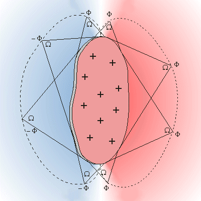
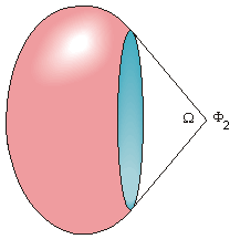
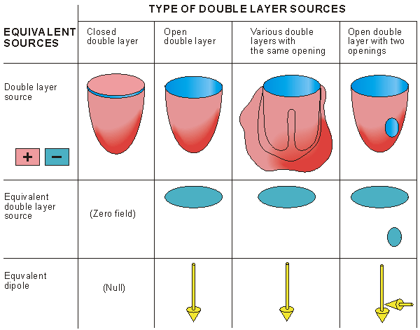
(11.7) Consider a closed uniform double layer. When such a double layer is seen from any point of observation, it can always be divided into two parts. One is seen from the positive side and the other is seen from the negative side, though each has exactly the same magnitude solid angle W, as described in Figure 11.3. (Double layer sources having more complex form can, of course, be divided into more than two parts.) These both produce a potential of the same magnitude, but because they have opposite signs, they cancel each other. As a result, a closed uniform double layer produces a zero field, when considered in its entirety.
Wilson et al. (1931) applied this principle to electrocardiography, since he understood the cardiac double layer source formulation. Suppose that the double layer formed by the depolarization in the ventricles includes a single wavefront, which is represented by a uniform double layer, and has the shape of a cup. If this cup is closed with a "cover" formed by a double layer of similar strength, then a closed surface is formed, that does not generate any potential field. From this we can conclude that the double layer having the shape of a cup can be replaced with a double layer having the shape of the cup's cover, but with its double layer oriented in the same direction as the cup, as described in Figure 11.4. From this example one can assert that two uniform double layers with the same periphery generate identical potential fields.
The field generated by a double layer disk at distances that are much greater than the disk radius appears to originate from a single dipole. In fact, at large enough distances from any dipole distribution, the field will appear to originate from a single dipole whose strength and orientation are the vector sum of the source components, as if they were all located at the same point. This is the reason why the electric field of the heart during the activation has a dipolar form and the concept of a single electric heart vector (EHV), as a description of the cardiac source, has a wide application. This is particularly true when the activation involves only a single ventricle. The true situation, where the right and left ventricle are simultaneously active, is more accurately represented by two separate dipoles.
This same argument may be used in explaining the effect of an infarct on the electric field of the heart. The infarct is a region of dead tissue; it can be represented by the absence of a double layer (i.e., an opening in a double layer). As a consequence, closing the double layer surface in this case introduces an additional cover, as shown in Figure 11.4. The latter source is a direct reflection of the effect of the infarct. (The paradox in this deduction is that the region of dead tissue is represented by an active dipole directed inward.)
Finally, we summarize the two important properties of uniform double layers defined by the solid angle theorem:
|
|
| ||||||
 |  |  | ||||||
|  | |||||||
W. T. Miller and D. B. Geselowitz (1978) developed a source model that is based directly on the generators associated with the activation of each cell. Their basic expression is patterned after Equation 8.23, which assigns a dipole source density to the spatial derivative of transmembrane voltage. For three dimensions, instead of a derivative with respect to a single variable, a gradient (including all three variables) is required. Consequently,
 i = -s i = -s Vm Vm | (11.8) |
| where | i | = dipole source density [µA/cm2] |
| s | = conductivity [mS/cm] | |
| Vm | = spatial derivative of transmembrane voltage [mV/cm] |
We examine the potential field at a point P, within or at the surface of a volume conductor, caused by a unit dipole
A similar expression holds for dipoles in the y and z directions.
where the coefficients cx, cy, and cz are found (as described above) by energizing the corresponding unit dipoles at point Q along x-, y-, and z-axes, respectively, and measuring the corresponding field potentials. Equations 11.9 and 11.10 are expressions of linearity, namely that if the source strength is increased by a factor c, the resultant voltage is increased by the same factor c. Since no other assumptions were required, Equation 11.10 is valid for any linear volume conductor, even for an inhomogeneous conductor of finite extent.
Fig. 11.5. Development of the lead vector concept.
Miller and Geselowitz used published data to evaluate action potential waveforms at various sites throughout the heart as well as times of activation. They could thus estimate Vm(x,y,z,t) and as a result, could evaluate the "actual" dipole moment per unit volume at all points. For simplicity the heart was divided into a finite number of regions, and the net dipole source strength in each region found by summing idV in that region.
In determining the surface potential fields the authors considered the number of dipole elements to be a small set (of 21) and evaluated the contribution from each. This part of their work constituted a relatively straightforward solution of the forward problem (dipole source in a bounded volume conductor). The reconstructed electrocardiograms showed very reasonable qualities.
11.4 LEAD VECTOR
11.4.1 Definition of the Lead Vector
PRECONDITIONS:
SOURCE: Dipole in a fixed location
CONDUCTOR: Finite (infinite), inhomogeneous
 (a unit vector in the x direction) in a fixed location Q, as illustrated in Figure 11.5. (Though the theory, which we will develop, applies to both infinite and finite volume conductors, we discuss here is only finite volume conductors, for the sake of clarity.)
Suppose that at the point P the potential FP due to the unit dipole is cx. (The potential at P must be evaluated relative to another local point or a remote reference point. Both choices are followed in electrophysiology, as is explained subsequently. For the present, we assume the existence of some unspecified remote reference point.) Because of our linearity assumption, the potential FP corresponding to a dipole px of arbitrary magnitude px is
(a unit vector in the x direction) in a fixed location Q, as illustrated in Figure 11.5. (Though the theory, which we will develop, applies to both infinite and finite volume conductors, we discuss here is only finite volume conductors, for the sake of clarity.)
Suppose that at the point P the potential FP due to the unit dipole is cx. (The potential at P must be evaluated relative to another local point or a remote reference point. Both choices are followed in electrophysiology, as is explained subsequently. For the present, we assume the existence of some unspecified remote reference point.) Because of our linearity assumption, the potential FP corresponding to a dipole px of arbitrary magnitude px is
FP = cx px(11.9) The linearity assumption ensures that the principle of superposition holds, and any dipole can be resolved into three orthogonal components px, py , pz
, pz , and the potentials from each superimposed. Thus we can express the potential FP at point P, due to any dipole at the point Q
FP = cx px + cy py + cz pz(11.10) 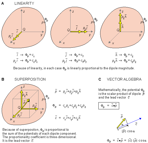 (A) Because of linearity, the potential at a point P in the volume conductor is linearly proportional to dipoles in each coordinate direction.
(B) By superposition the potential at the point P is proportional to the sum of component dipoles in each coordinate direction. This proportionality is three-dimensional and can therefore be considered as a vector
(A) Because of linearity, the potential at a point P in the volume conductor is linearly proportional to dipoles in each coordinate direction.
(B) By superposition the potential at the point P is proportional to the sum of component dipoles in each coordinate direction. This proportionality is three-dimensional and can therefore be considered as a vector  , called lead vector.
(C) The potential at the point P is the scalar product of the source dipole and the lead vector . Equation 11.10 can be simplified if the coefficients cx, cy, and cz are interpreted as the components of a vector . This vector is called the lead vector. Consequently, Equation 11.10 can be written
, called lead vector.
(C) The potential at the point P is the scalar product of the source dipole and the lead vector . Equation 11.10 can be simplified if the coefficients cx, cy, and cz are interpreted as the components of a vector . This vector is called the lead vector. Consequently, Equation 11.10 can be written
FP = ·  | (11.11) |
The lead vector is a three-dimensional transfer coefficient which describes how a dipole source at a fixed point Q inside a volume conductor influences the potential at a point within or on the surface of the volume conductor relative to the potential at a reference location. The value of the lead vector depends on:
We tacitly assume that the potential at the reference is zero and hence does not have to be considered. Note that the value of the lead vector is a property of the lead and volume conductor and does not depend on the magnitude or direction of the dipole .
It can be shown that in an infinite, homogeneous volume conductor the lead vector is given by the sum of components along lines connecting the source point with each of the two electrode points (each scaled inversely to its physical length). The same also holds for a spherical, homogeneous volume conductor, provided that the source is at the center.
For each location P0 . . . Pn of P, that lies within or at the surface of the volume conductor, we can determine a lead vector 0 . . . n for the dipole at a fixed location, so that, according to Equation 11.11, we have
| Fi = i · | (11.12) |
Then the potential difference between any two points Pi and Pj is
| Vi j = Fi - Fj | (11.13) |
This describes the voltage that would be measured by the lead whose electrodes are at Pi and Pj. To what lead vector does this lead voltage correspond? Consider first the vector ij formed by
| i j = i - j | (11.14) |
Now the voltage between the points Pi and Pj given by Equation 11.13 can also be written, by substitution from Equation 11.12, as follows:
| Vi j = Fi - Fj = i · - j · = i j · | (11.15) |
hence identifying i j as the lead vector for leads Pi - Pj. From this result we can express any bipolar lead voltage V as
 | (11.16) |
where is a lead vector. We note that Equation 11.16 for bipolar leads is in the same form as Equation 11.11 for monopolar leads. But Equations 11.14-11.16 can be interpreted as that we may first determine the lead vectors i and j corresponding to unipolar leads at Pi and Pj, respectively, and then form their vector difference, namely ij. Then the voltage between the points Pi and Pj, as evaluated by a bipolar lead, is the scalar product of the vector ij and the dipole , as shown in Figure 11.6 and described by Equation 11.16.
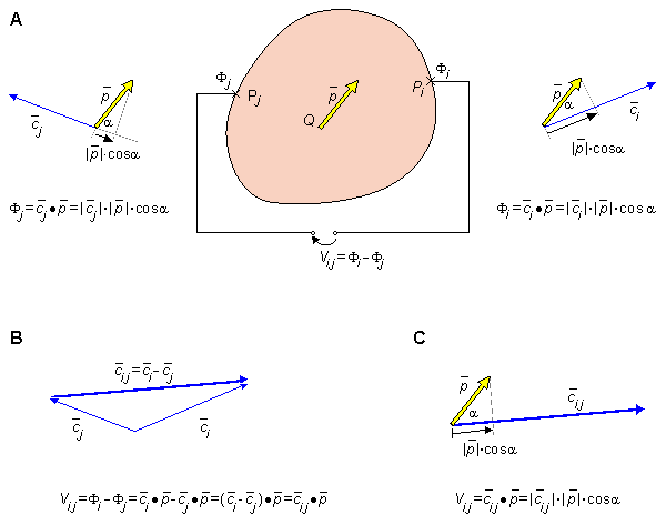
Fig. 11.6. Determination of the voltage between two points at or within the surface of a volume conductor.
(A) The potentials Fi and Fj at Pi and Pj due to the dipole may be established with scalar products with the lead vectors i and j , respectively.
(B) For determining the voltage Vi j between Pi and Pj, the lead vector i j = i - j is first determined.
(C) The voltage Vi j is the scalar product of the lead vector i j and the dipole .
In Einthoven's electrocardiographic model the cardiac source is a two-dimensional dipole in a fixed location within a volume conductor that is either infinite and homogeneous or a homogeneous sphere with the dipole source at its center.
Einthoven defined the potential differences between the three pairs of these three points to constitute the fundamental lead voltages in electrocardiography. These are designated VI, VII, and VIII and are given by
Since
For the lead vectors we obtain:
Ernest Frank measured the lead vectors of the scalar leads by constructing an electrolytic tank model of the human torso (Frank, 1954). The following values were obtained for the three lead vectors of the standard leads. Note that only the relative values of these lead vectors have any meaning because the measurement procedure was not calibrated.
We noted earlier that since VI + VIII = VII, a condition dictated by Kirchhof's law, the corresponding lead vectors must form a closed triangle. One can confirm from Equation 11.21 that, indeed,
Lead vector concept was first introduced by H. C. Burger and J. B. van Milaan (1946, 1947, 1948) (Burger, 1967), who also used an inhomogeneous electrolyte tank model of the human torso to measure the lead vectors of standard leads.
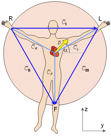
Einthoven first recognized that because the limbs are generally long and thin, no significant electrocardiographic currents from the torso would be expected to enter them. Accordingly, Einthoven realized that the potential at the wrist was the same as at the upper arm, while that at the ankle was the same as at the upper thigh. Einthoven consequently assumed that the functional position of the measurement sites of the right and left arm and the left leg corresponded to points on the torso which, in turn, bore a geometric relationship approximating the apices of an equilateral triangle. He further assumed that the heart generator could be approximated as a single dipole whose position is fixed, but whose magnitude and orientation could vary. The location of the heart dipole relative to the leads was chosen, for simplicity, to be at the center of the equilateral triangle. (In matter of fact, the Einthoven assumptions and model were not truly original, but were based on the earlier suggestions of Augustus Waller (1889).)
Because of the central location of the heart dipole in the Einthoven model, the relationship between potentials at the apices of the triangle are the same whether the medium is considered uniform and infinite in extent, or the volume conductor is assumed to be spherical and bounded. For the unbounded case, we can apply Equation 8.12, which may be written FP = ·  r /(4psr 2 ) from which we learn that the lead vector for a surface point P is r /(4psr 2 ) - that is, along the radius vector to P. Point P is, according to Einthoven, at the apices of the equilateral triangle. Consequently, if the right and left arms and left foot are designated R, L, and F, respectively, then the three corresponding lead vectors R, L, and F are the radius vectors between the origin and the corresponding points on the equilateral triangle, as illustrated in Figure 11.7. From the aforementioned, the potentials at these points are:
FR = R · FL = L · (11.17) FF = F ·
VI = FL - FR = L · - R · = (L - R ) · = I · VII = FF - FR = F · - R · = (F - R ) · = II · (11.18) VIII = FF - FL = F · - L · = (F - L ) · = III · R, L, and F are equal in magnitude and each is in the direction from the origin to an apex of the equilateral triangle, then I, II, and III must lie along a leg of the triangle (since I = L - R, etc.) For example I is seen to lie oriented horizontally from the right arm to the left arm.
In summary, VI, VII, and VIII are the three standard limb leads (or scalar leads) in electrocardiography. From Equation 11.18 one can confirm that the three lead vectors I, II, and III also form an equilateral triangle, the so-called Einthoven triangle, and these are shown in Figure 11.7.
The limb lead voltages are not independent, since VI + VIII - VII = 0 , as can be verified by substituting for the left side of this equation the component potentials from Equation 11.18, namely (FL - FR) + (FF - FL) - (FF - FR), and noting that they do, in fact, sum to zero. The above relationship among the standard leads is also expressed by I· + III· - II· = 0, according to Equation 11.18. Since is arbitrary, this can be satisfied only if I + III - II = 0, which means that the lead vectors form a closed triangle. We were already aware of this for the Einthoven lead vectors, but the demonstration here is completely general.

(11.19) 
I = II = 0.5 - 0.87(11.20) III = - 0.5 - 0.87 Frank Triangle
PRECONDITIONS:
SOURCE: (Three-dimensional) dipole in a fixed location
CONDUCTOR: Finite, homogeneous
I = - 14 + 76 + 27 II = 16 + 30 - 146(11.21) III = 30 - 46 - 173 I + II - III = 0 and hence form a closed triangle. This triangle is called the Frank triangle, and it is illustrated in Figure 11.8.
Burger Triangle
PRECONDITIONS:
SOURCE: Dipole in a fixed location
CONDUCTOR: Finite, inhomogeneous
The lead vectors, which they measured, are given below. Since these vectors must necessarily form a closed triangle (just as Einthoven and Frank triangles), this triangle has been called Burger triangle; it is shown in Figure 11.8. The absolute values of the lead vectors have no special meaning since no calibration procedure was carried out. The lead vectors obtained were
I = - 17 + 65 + 21 II = 15 + 25 - 120(11.22) III = 32 - 40 - 141 We may compare the three triangles described so far (i.e., the Einthoven, Frank, and Burger) by normalizing the y-component of each I vector to 100. This means that the values of the Einthoven triangle components must be multiplied by 100, those of the Frank triangle by 100/76 = 1.32, and those of the Burger triangle by 100/65 = 1.54. (The reader can confirm that in each case I = 100 results.) The resulting lead vector components are summarized in Table 11.1.
One may notice from the table that in the measurements of Frank and Burger, the introduction of the boundary of the volume conductor has a great influence on the lead vectors. As pointed out earlier, the lead vector also depends on the dipole location; thus these comparisons may also reflect differences in the particular choice that was made. Figure 11.8 illustrates the Einthoven, Frank, and Burger triangles standardized according to Table 11.1.
| Lead | Triangle | cx | cy | cz |
| cI | Einthoven Frank Burger | -18 -26 | 100 100 100 | 36 32 |
| cII | Einthoven Frank Burger | 21 23 | 50 40 38 | -87 -192 -185 |
| cIII | Einthoven Frank Burger | 39 49 | -50 -61 -62 | -87 -228 -217 |
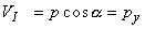
The shape of the Frank and Burger triangles was recently investigated by Hyttinen et al. (1988). Instead of evaluating the lead vectors for a single dipole location, they examined the effect of different dipole positions within the heart. According to these studies the shape of the Frank and Burger triangles varies strongly as a function of the location of the assumed heart dipole . They showed that the difference between the original Frank and Burger triangles is not necessarily so small if the dipole is placed at other locations. Figures 11.9 and 11.10 illustrate the variation of the Frank and Burger triangles as functions of the source location. Tables 11.2A and 11.2B compare the lead vectors for the Einthoven, Frank, and Burger triangles from two source locations.
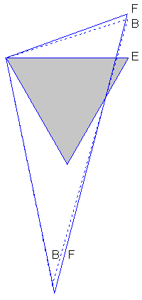
Fig. 11.8. Einthoven (E), Frank (F), and Burger (B) triangles. Note that the Einthoven triangle lies in the frontal plane, whereas the Frank and Burger triangles are tilted out of the frontal plane. Only their frontal projections are illustrated here.
Fig. 11.9. Variation of the Frank triangle as a function of dipole location. The black circle in the miniature lead vector triangles arising from the Frank torso are superimposed on the site of the dipole origin. (From Hyttinen et al., 1988.).
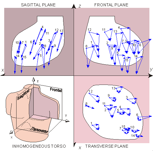
Fig. 11.10. Variation of the Burger triangle as a function of source location. (From Hyttinen et al., 1988.).
| Lead | Triangle | cx | cy | cz |
| cI | Einthoven Frank Burger | -2.8 -31.2 | 100 100 100 | -1.8 -6.4 |
| cII | Einthoven Frank Burger | 16 97 | 50 53 46 | -87 -88 -162 |
| cIII | Einthoven Frank Burger | 19 135 | -50 -47 -57 | -87 -86 -163 |
| Lead | Triangle | cx | cy | cz |
| cI | Einthoven Frank Burger | -6.6 -3.0 | 100 100 100 | 12 -8.2 |
| cII | Einthoven Frank Burger | 23 33 | 50 44 62 | -87 -117 -217 |
| cIII | Einthoven Frank Burger | 30 30 | -50 -60 -39 | -87 -130 -209 |
For a fixed-source dipole lying within a given volume conductor, the lead vector depends solely on the location of the field point. A lead vector can be found associated with each point on the volume conductor surface. The tips of these lead vectors sweep out a surface of its own. This latter surface is known as the image surface.
One could, in principle, consider a physical surface lying within a volume conductor of finite or infinite extent and evaluate an image surface for it in the same way as described above. However, most interest is concentrated on the properties of fields at the bounding surface of volume conductors, since this is where potentials are available for noninvasive measurement. Consequently, the preconditions adopted in this section are for a dipole source (multiple sources can be considered by superposition) lying in a bounded conducting region.
We accept, without proof, that any physical volume conductor surface has an associated image surface for each dipole source location. This seems, intuitively, to require only that no two points on the physical surface have the same lead vector - a likely condition for convex surfaces. The image surface is a useful tool in characterizing the properties of the volume conductor, such as the effect of the boundary shape or of internal inhomogeneities, independent of the effect of the leads. That is, one could compare image surfaces arising with different inhomogeneities without having to consider any particular lead system.
A simple example of an image surface is given by a uniform spherical volume conductor with dipole source at its center. We have seen that for this situation the unipolar lead vector is proportional to the radius vector from the center of the sphere to the surface field point. Therefore, the image surface for a centric source in a uniform sphere is also a sphere.
We now describe how to construct the image surface for any linear volume conductor of arbitrary shape. It is done by placing a unit dipole source at a chosen point within the conductor in the direction of each coordinate axis and then measuring the corresponding potential at every point on the surface. For the unit vector along the x-axis, the potentials correspond precisely to the lead vector component in the x direction, as is clear from Equation 11.10. Similarly for the y and z directions, and therefore, the lead vectors can be determined in space from these measurements, and they form the image surface for the chosen source location. This procedure and the resulting image surface are illustrated in Figure 11.11.
Note that the shadings in Figures 11.11, 11.12, and 11.13 are not arbitrary; rather, they illustrate that for the region inside the volume conductor, the corresponding region in the image space is farther from the origin..
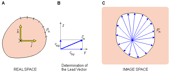
Fig. 11.11. Construction of the image surface for a source point at a volume conductor of arbitrary shape, illustrated in two dimensions. A one-to-one relation is established between points on the surface of the volume conductor and the image surface.
(A) Unit vectors are placed at the source location.
(B) By measuring the corresponding potentials at each surface point, the lead vector can be determined.
(C) The locus described by the family of lead vectors form the image surface.
 | (11.23) |
From Figure 11.12 it is easy to see the following relationship between the lead vectors 1, 2, and s:
 | (11.24) |
Therefore, the voltage, measured in the real space from the point P must fulfill the requirement:
 | (11.25) |
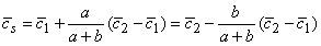
The point that fulfills this requirement in the real space can be found in the following way: We connect between the points P1 and P2 two resistors in series having the resistance ratio of a/b. The point P is at the interconnection of these resistors. (We must choose Ra and Rb large enough so that the current through this pathway has a negligible effect on F1 and F2.)
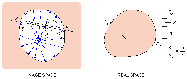
Fig. 11.12. Determination of the point P in the real space corresponding to an image space point P' located inside the image surface.
To begin, we construct the image surface of the volume conductor in relation to the known location of the dipole. Now we want to find two points on the surface of the volume conductor such that the voltage between them is proportional only to the y-component of the dipole. Mathematically, this can be formulated as:
| V21 = 21 · = c21x px + c21y py + c21z pz | (11.26) |
and we seek a lead vector 21, which both lies in the image space and is oriented solely in the direction of the y-axis. This corresponds to identifying any pair of points on the image surface that are located at the intersections of a line directed parallel to the y-axis. The voltage measured between those points in real space is consequently proportional only to the y-component of the dipole. To obtain the largest possible signal (in order to minimize the noise), we select from all image space points that fulfill the requirement discussed above, the one with the longest segment (maximum lead vector), as illustrated in Figure 11.13.
If we want to measure all three orthogonal components of the dipole source, we repeat this procedure for the z and x directions. Because the resulting (maximum) lead vectors are usually not of equal length, we equalize the measured signals with a resistor network to obtain both an orthogonal and a normalized lead system. Such a normalizing procedure is described in Figure 11.13 for a two-dimensional system. In this case two resistors, RA and RB, form a simple voltage-divider, and the output voltage is reduced from the input by RB/(RA + RB). We choose this ratio to compensate for a lead vector amplitude that is too large. Note that the assumed voltage-divider behavior requires that the voltage-measuring circuit (amplifier) has a sufficiently high input impedance for negligible loading..
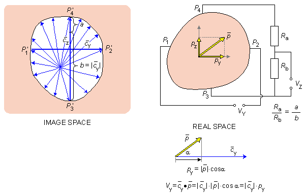
34 for surface points P3 - P4 is solely in the z direction, whereas lead 12 established for surface points P1 - P2 is solely in the y direction. Since |34|/|12| = (a + b)/b, the resistor network Ra and Rb is inscribed to reduce the voltage from (P3 - P4) by b/(a + b), hence making the effective z-lead equal to the y-lead in magnitude.Frank adopted the following coordinate system for the model: The model was divided into 12 levels with horizontal planes at increments of 5 cm (2 inches). The center of the heart was located on level 6 about 4 cm to the front of the plane located at the midline of the right and left arms, and about 2.5 cm to the left of the sagittal plane located at the midline of the model. On each horizontal plane, 16 points were established by drawing 8 lines through the midline of the model (within increments of 22.5°). The intersections of these lines on the surface of the model were labeled with letters A through P in a clockwise direction starting from the left side, as shown in Figure 11.14. Note that the coordinate axis nomenclature used here is not the same as that adopted by Frank, since the consistent coordinate system of the Appendix A has been used.
Figures 11.15, 11.16, and 11.17 illustrate the image surface measured by Frank in the three projections - the frontal, sagittal, and transverse planes. The figures also show the points corresponding to the Einthoven limb leads, which in this case form the Frank triangle.
The concept of lead field is a straightforward extension of the concept of lead vector. In the evaluation of a lead field, one follows a procedure that is just the reverse of that followed in obtaining the image surface. These may be contrasted as follows (see Figure 11.19).
The source was a dipole The measurement point was varied over the surface of the volume conductor (Figure 11.11A).
The image surface was generated by the tips of the lead vectors We assume a fixed electrode pair defining a lead (fixed measurement sites).
We observe the behavior of the lead vector We assign
With this latter procedure, it is possible to evaluate the variation of the lead vector With the concept of lead field it is possible both to visualize and to evaluate quantitatively the sensitivity distribution of a lead within a volume conductor, since it is the same as the field of a reciprocal current.
The actual measurement of sensitivity distribution (using either a torso-shaped tank model or a computer model) can be accomplished more easily using reciprocity.
Because the reciprocal current corresponds to the stimulating current introduced by a lead in electric stimulation, they have exactly the same distribution.
The sensitivity distribution in the measurement of electric impedance of the tissue may be similarly determined with the concept of lead field.
Because the principle of reciprocity and the concept of lead field are valid also in magnetic fields, all of these points are true for the corresponding magnetic methods as well.
Furthermore, the concept of lead field easily explains the similarities and differences in the sensitivity distributions between the corresponding electric and magnetic methods.
Fig. 11.19. The definition of the lead field and different ways to illustrate it.
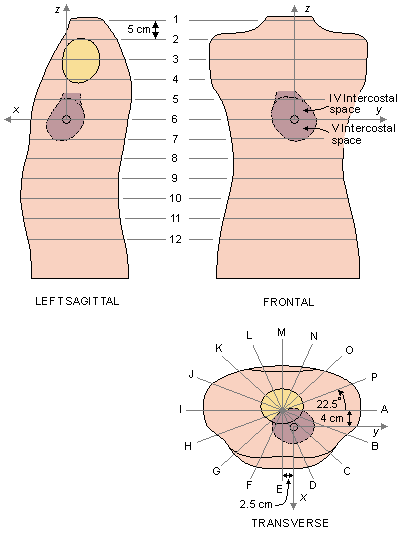
Fig. 11.14. The Frank torso model and coordinate system. (The latter has been related to correspond with the system adopted in this text and discussed in the Appendix A.).
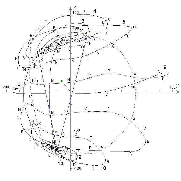
Fig. 11.15. The image surface of the Frank torso model in frontal view.
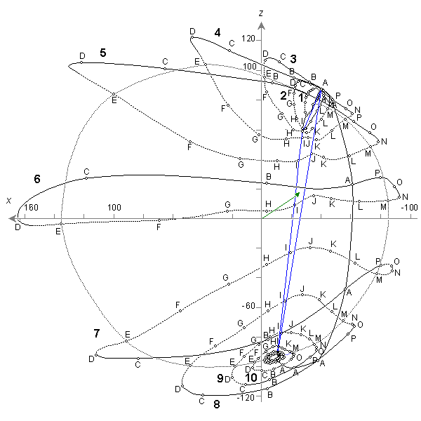
Fig. 11.16. The image surface of the Frank torso model in sagittal view.
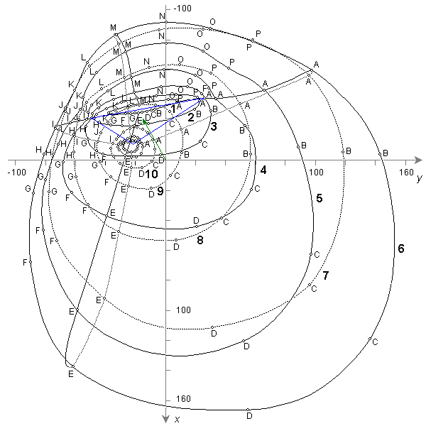
Fig. 11.17. The image surface of the Frank torso model in transverse view.
11.5.6 Recent Image Surface Studies
In recent years the image surface for the human torso has been investigated using computer models. In these models one can introduce not only the effect of body shape but also inhomogeneities such as the lungs, intracavitary blood masses, surface muscle layers, and so on. One such study is that of Horácek (1971), who included the effect of body shape, lungs, and intracavitary blood. Horácek observed that the lungs and the intracavitary blood masses can substantially distort the image surface and consequently cause variations in the body surface potential distribution. However, because of the complexity of the effect, no simple universal statement can be made to describe the influence of the inhomogeneities.
A modified Horácek model that includes the skeletal muscle was developed and studied by Gulrajani and Mailloux (1983). The latter authors chose to examine the effects of modifications introduced by inhomogeneities in terms of effects on body surface potentials rather than on the image surface per se.
11.6 LEAD FIELD
11.6.1 Concepts Used in Connection with Lead Fields
It is useful to start a discussion of the lead field by first introducing the concept of sensitivity distribution. As noted in Section 11.4.3, the lead vector has different values for different source locations. In other words, for a given field point, the length and direction of the lead vector vary as a function of the source location. For a fixed field point location, one can assign to each possible source point the value of the lead vector. In this way we establish a lead vector field, which is distributed throughout the volume conductor. Because the lead vector indicates the sensitivity of the lead to the dipole source through V = · (Equation 11.16), the distribution of the magnitude and the direction of the lead vector is at the same time the distribution of the sensitivity of the lead to the dipole source as a function of its location and orientation. This is further illustrated in Figure 11.18. (It should be emphasized that the concept of sensitivity distribution is not limited to the detection of bioelectric sources. The same concept is applicable also to the measurement of tissue impedance.)
For later use we will define the concepts of isosensitivity surface or isosensitivity line and half-sensitivity volume. An isosensitivity surface is a surface in the volume conductor, where the absolute value of the sensitivity is constant. When sensitivity distributions are illustrated with two-dimensional figures, the isosensitivity surface is illustrated with isosensitivity line(s). The concept of isosensitivity surface is used to enhance our view of the distribution of the magnitude of the sensitivity. The isosensitivity surface where the absolute value of the sensitivity is one half of its maximum value within the volume conductor separates a volume called half-sensitivity volume. This concept can be used to indicate how concentrated the detector's sensitivity distribution is..
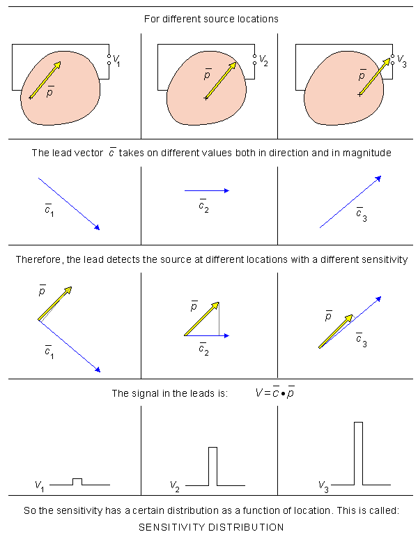
Fig. 11.18. The concept of sensitivity distribution.
11.6.2 Definition of the Lead Field
PRECONDITIONS:
SOURCE: Volume source
CONDUCTOR: Finite (infinite), inhomogeneous
In our definition of the image surface (Section 11.5.1):
located at a fixed point.
associated with all surface sites (Figure 11.11C).
In evaluating the lead field we proceed the other way around:
as a function of the location of the dipole source varying throughout the volume conductor (Figure 11.19A)
to the location of (which for a volume source is a field of dipole elements k ).
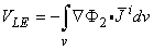
within the volume conductor. This field of lead vectors is called the lead field L, as noted earlier and illustrated in Figure 11.19A. Therefore, the lead field theory applies to distributed volume sources. The procedure may be carried out with a finite or an infinite volume conductor. In any physically realizable system, the volume conductor is necessarily finite, of course. Thus the preconditions for the discussion on the lead field are those defined above.
From the behavior of the lead vector as a function of the location k of the dipole source , we can easily determine the lead voltage VL generated by a distributed volume source (see Figure11.19B). The contribution Vk of each elementary dipole k to the lead voltage is obtained, as was explained in Section 11.4.1, with Equation 11.16 by forming the scalar product of the dipole element k and the lead vector k at that location, namely Vk = k · k. The total contribution of all dipole elements - that is, the total lead voltage - is, according to the principle of superposition, the sum of the contributions of each dipole element k , namely
VL = S k · k . Mathematically this will be described later by Equations 11.30 and 11.31, where the dipole element k is replaced by the impressed current source element idV (where i has the dimensions of dipole moment per unit volume).
The lead field has a very important property, which arises from the reciprocity theorem of Helmholtz. It is that for any lead, the lead field LE is exactly the same as the current flow field resulting from the application of a unit current I r , called the reciprocal current, to the lead (Figure 11.19C). In this procedure the lead is said to be reciprocally energized. It is this correspondence that makes the lead field concept so very powerful in the following way:
The lead field may be visualized either as a field of lead vectors, as in Figure 11.19C, or with lead field current flow lines, as in Figure 11.19D. The relationship between these two methods is, obviously, that the lead vectors are tangents to the lead field current flow lines and that their length is proportional to the density of the flow lines. The reciprocity theorem is further discussed in the next section in greater detail.
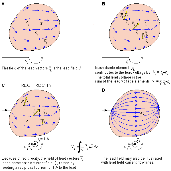 (A) When defining the lead field, we assume a fixed electrode pair constituting a lead, and we observe the behavior of the lead vector as a function of the location k of the dipole source within the volume conductor. This field of lead vectors is the lead field L.
(B) When we know the lead vector at each location k, we obtain the contribution of each dipole element k to the lead voltage: Vk = k · k . Due to superposition, the total lead voltage VL is the sum of the lead voltage elements.
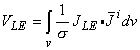
(C) Based on the reciprocity theorem, the lead field LE is the same as the electric current field if a (reciprocal) current I r of 1 A is introduced to the lead. The lead voltage due to a volume source of distribution i is obtained through integrating the dot product of the lead field current density and the source density throughout the volume source.
(D) The lead field may also be illustrated with the lead field current flow lines.
(A) When defining the lead field, we assume a fixed electrode pair constituting a lead, and we observe the behavior of the lead vector as a function of the location k of the dipole source within the volume conductor. This field of lead vectors is the lead field L.
(B) When we know the lead vector at each location k, we obtain the contribution of each dipole element k to the lead voltage: Vk = k · k . Due to superposition, the total lead voltage VL is the sum of the lead voltage elements.
(C) Based on the reciprocity theorem, the lead field LE is the same as the electric current field if a (reciprocal) current I r of 1 A is introduced to the lead. The lead voltage due to a volume source of distribution i is obtained through integrating the dot product of the lead field current density and the source density throughout the volume source.
(D) The lead field may also be illustrated with the lead field current flow lines.11.6.3 Reciprocity Theorem: the Historical Approach
The lead field theory that is discussed in this section is based on a general theory of reciprocity, introduced by Hermann von Helmholtz in 1853 (Helmholtz, 1853). Its application to the formulation of lead field theory was carried out 100 years later by Richard McFee and Franklin D. Johnston (1953, 1954,ab) as well as by Robert Plonsey (1963) and by Jaakko Malmivuo (1976). Before describing the lead field theory in more detail, we consider first the reciprocity theorem of Helmholtz.
Though Helmholtz introduced the principle of reciprocity in connection with bioelectricity, it is a general property of linear systems, not limited only to bioelectricity. Helmholtz described the principle of reciprocity, in its original form, with the following example, which, it should be noted, also includes (for the first time) the principle of superposition.
A galvanometer is connected to the surface of the body. Now every single element of a biological electromotive surface produces such a current in the galvanometer circuit as would flow through that element itself if its electromotive force were impressed on the galvanometer wire. If one adds the effects of all the electromotive surface elements, the effect of each of which are found in the manner described, he will have the value of the total current through the galvanometer.
In other words, it is possible to swap the location of the (dipole) source and the detector without any change in the detected signal amplitudes. (Note that Helmholtz used a voltage double layer source and measured the current produced by it, whereas in our case the source is considered to be a current dipole or a collection of dipoles such as implied in a double layer source, whereas the measured signal is a voltage.)
Helmholtz illustrated the leading principle of the reciprocity theorem with the following example, described in Figure 11.20. This example includes two cases: case 1 and case 2.
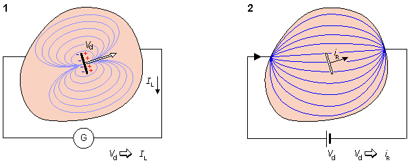
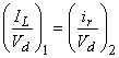
Fig. 11.20. Illustration of the reciprocity theorem of Helmholtz.
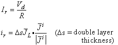
We first consider case 1: A galvanometer (i.e., an electric current detector) G is connected at the surface of the volume conductor. Inside the conductor there is a differential element of double layer source, whose voltage is Vd and which causes a current IL in the galvanometer circuit.
We now consider case 2: The double layer source element is first removed from the volume conductor. Then the galvanometer is replaced by an electromotive force of the same magnitude Vd as the voltage of the double layer source. This produces a reciprocal current ir through the same differential area at the (removed) double layer source element in the volume conductor.
Now the reciprocity theorem of Helmholtz asserts that the current IL flowing in case 1 through the galvanometer is equal to the current ir flowing in case 2 through the differential area located at the (removed) double layer source element. This result is expressed in equation form as:
 | (11.27) |
where the left-hand side of the equation denotes case 1 and the right-hand side case 2.
If we make the area of the double layer K times larger, the current IL through the galvanometer in case 1 is now (by the application of superposition) K times larger - that is, KIL. In case 2, the electromotive force Vd in the galvanometer wire remains the same, because it represents the (unchanged) voltage over the double layer source in case 1. Therefore, it still produces the same current density in the source area. But because the source area is now K times larger, the total current through it is also K times larger - that is, Kir. Consequently, Equation 11.27 becomes
 | (11.27A) |
and dividing both sides by K returns it to the expression arising from the original area. (In the above one should keep in mind that the original area A and KA are assumed to be very small so that ir and Vd can be considered uniform.)
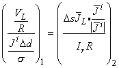
i, whereas the signal is the lead voltage VL produced by it. We can directly obtain these expressions from those of Helmholtz by application of the principle of duality. The result, illustrated in Figure 11.21, is discussed below..
| |
 |  |
|
|
Fig. 11.21. Derivation of the equation for lead field theory.
Since Helmholtz's theorem applies to a discrete source, we make the following assumptions:
The lateral extent of the voltage double layer element Vd is differential - that is, Ds.
The separation of the poles of the corresponding dipole element The conductivity at the source point is s.
The resistance of the galvanometer circuit between the measurement points equals R.
iDs is Dd, where i is an applied current density so that iDd has the dimensions of a double layer source.
In case 1 and case 2 we may further evaluate the following expressions. For case 1:
Instead of the current IL measured by the galvanometer, we can examine the related lead voltage VL = RIL, or IL = VL/R. (To prevent the galvanometer from affecting the volume conductor currents and voltages in a real situation, R should be chosen, but this choice does not affect the validity of this expression.)
Instead of reference to a voltage source Vd, we now emphasize the concomitant current source i = Vds/Dd (Equation 11.2), where by rearranging we have Vd = iDd/s.
For case 2:
Instead of examining the reciprocal current density ir at the (removed) source point we can evaluate the related lead field current density The required voltage source Vd in the circuit connected to the conductor can be achieved if we use a reciprocal current source Ir = Vd/R, since then we have Vd = IrR.
L = ir/Ds. These are connected by ir = Ds · L. The dot product is required here because the current ir is the component of the reciprocal current flowing through the source area in the direction of the source i. This can also be written
ir = DsL · i/|i|.
Substituting these equivalencies into the equation of Helmholtz, namely Equation 11.27, we obtain:
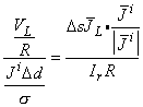 | (11.28) |
where the left-hand side of the Equation 11.28 denotes case 1 and the right-hand side case 2 in the Helmholtz procedure, respectively. Solving for the lead voltage VL in Equation 11.28, we obtain
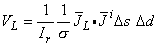 | (11.29) |
where Ds Dd = Dv, which is the volume element of the source. (In the limit, Dv
where
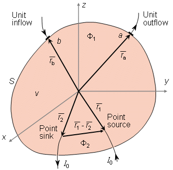
If we subtract the second equation from the first one, integrate term by term over the volume v, and use the divergence theorem, we obtain
Since
(Thus IF is a flow source, as defined earlier in Equation 8.35.) We assume further that F2 is the scalar potential produced solely by current caused to cross the surface S with a current density J [A/m2]. Usually we assume that J flows from conducting electrodes of high conductivity compared with s, so that the direction of J is normal to the bounding surface. In this case J can be specified as a scalar corresponding to the flow into v. (The scalar potential F2 is later identified as the reciprocal electric scalar potential due to the reciprocal current Ir fed to the lead.) Since the current J is solenoidal, it satisfies
The scalar fields F1 and F2 satisfy the following equations:
since IF is a source of F1 and
since the field F2 is established by the applied current J.
For the source J at the surface, the current must be solenoidal everywhere in v; hence:
We may rewrite Equation 11.33 by substituting Equations 11.38 and 11.37 into its left-hand side, and Equations 11.38 and 11.36 into its right-hand side, obtaining
which is the desired form of the reciprocity theorem.
where
where
If we choose I0 to be unity, then this equation shows that the voltage between two arbitrary surface points a and b due to a unit current supplied internally between points 1 and 2 equals the voltage between these same points 2 and 1 due to a unit current applied externally (reciprocally) between points a and b. This is essentially the reciprocity theorem of Helmholtz.
dv.) By extending Equation 11.29 throughout all source elements, and choosing the reciprocal current to be a unit current Ir = 1 A, we may write:
(11.30) LE denotes an electric lead field due to unit reciprocal current. Note that although i was originally defined as a current density, it may also be interpreted as a volume dipole density, as is clear in Equation 11.30 and by their similar dimensions. Equation 11.30 is the most important equation in the lead field theory, as it describes the lead voltage (the electric signal in a lead) produced by an arbitrary volume source described by i(x,y,z). It may be stated in words as follows:
To determine the lead voltage produced by a volume source, we first generate the lead field in the volume conductor by feeding a unit (reciprocal) current to the lead. Every element of the volume source contributes to the lead voltage a component equal to the scalar product of the lead field current density and the volume source element divided by the conductivity.
If the volume conductor is homogeneous throughout the source region, we may move the coefficient 1/s outside the integral and write:
(11.31) According to Equation 11.31, the lead field has an important property: it equals the lead sensitivity distribution. This means that at each point of the volume conductor, the absolute value of the lead field current density equals to the magnitude of the lead sensitivity, and the direction of the lead field current equals the direction of the lead sensitivity. It should be noted that the lead field fully takes into account the effect of the volume conductor boundary and internal inhomogeneities; hence these have an effect on the form of the lead field. (The concept of secondary sources is contained within lead field theory through the effect of the inhomogeneities on the form of the lead field.)
Lead field theory is a very powerful tool in bioelectromagnetism. It ties together the sensitivity distribution of the measurement of bioelectric sources, distribution of stimulation energy, and sensitivity distribution of impedance measurements, as is explained later. In general, if the lead and the volume conductor are known, the distribution of the lead sensitivity may be determined, based upon lead field theory. On the other hand, if the source and the volume conductor are known, the distribution of the actual field may be determined directly without using the lead field concept. All this holds for corresponding biomagnetic phenomena as well.
11.6.5 Field-Theoretic Proof of the Reciprocity Theorem
A brief explanation of Helmholtz's reciprocity theorem was given in Section 11.6.3, without offering a mathematical proof. That explanation was based on the ideas of the original publication of Helmholtz (1853). The field-theoretic proof of the reciprocity theorem as described by Plonsey (1963) is presented below.
Proof of the Reciprocity Theorem
Consider an arbitrary volume v bounded by the surface S and having a conductivity s (which may be a function of position). If F1 and F2 are any two scalar fields in v, the following vector identities must be satisfied:
· F1 (sF2 ) = F1 · (sF2 ) + sF1 · F2 (11.32) · F2 (sF1 ) = F2 · (sF1 ) + sF2 · F1

(11.33) F1 · F2 = F2 · F1 , these terms cancel out in deriving Equation 11.33 from 11.32. The derivation of Equation 11.33 is well known in the physical sciences; it is one of a number of forms of Green's theorem.
Now we assume that F1 is the scalar potential in volume v due to sources within it specified by the equation
IF = - · i(11.34)

(11.35)
· (sF1 ) = - IF(11.36)
sF2 · d = J dS(11.37) Since -sF2 carries the direction of the current (= L ) and d is the outward surface normal, Equation 11.37 shows that for our chosen signs J is positive for an inflow of current. No current due to the source IF crosses the boundary surface (since in this case it is totally insulating), and hence
F1 · d = 0(11.38)
· (sF2) = 0(11.39)
(11.40) The Reciprocity Theorem of Helmholtz
The reciprocity theorem of Helmholtz can be derived from Equation 11.40 in the following way. Consider that F2 (the reciprocal electric scalar potential) arises from a particular distribution J, where an inflow of a unit (reciprocal) current is concentrated at point  b on the surface and an outflow of a unit (reciprocal) current at point a on the surface, where a and b are position vectors shown in Figure 11.22. Note that this is opposite to the current flow direction given in Plonsey (1963). (With this sign notation, current dipoles in the direction of the lead field current produce a positive signal in the lead, as will be seen later in Equation 11.50 (Malmivuo, 1976).)
The above can be expressed mathematically:
b on the surface and an outflow of a unit (reciprocal) current at point a on the surface, where a and b are position vectors shown in Figure 11.22. Note that this is opposite to the current flow direction given in Plonsey (1963). (With this sign notation, current dipoles in the direction of the lead field current produce a positive signal in the lead, as will be seen later in Equation 11.50 (Malmivuo, 1976).)
The above can be expressed mathematically:
J =  s(b - ) - s(a - )
(11.41) s is a two-dimensional unit Dirac delta function on the bounding surface, and hence the magnitude of both inflow and outflow is unity.
Consider IF to consist of a point source of current I0 at 1 and a point sink of equal magnitude at 2, where 1 and 2 are the position vectors shown in Figure 11.22. Now IF can be written:
IF = I0[v(1 - ) - v(2 - )](11.42) v is a three-dimensional Dirac delta function.
Substituting Equations 11.41 and 11.42 into Equation 11.40, we obtain

(11.43)
2 by means of a Taylor series expansion:
| F2 (1) = F2 (2) + F2 · (1 - 2) + . . . | (11.44) |
Note that since the field F2 is established by currents introduced at the surface into a source-free region, it is well behaved internally and a Taylor series can always be generated. If we let (
Denoting the voltage between the points a and b as
and substituting Equations 11.45 and 11.46 into Equation 11.43, we obtain
Because the impressed current sources are totally contained within S, the integrand is zero everywhere on S, and the first term on the right-hand side of Equation 11.49 is zero; thus we obtain
The quantity -
where
This reciprocal current field s is defined as the lead field.
If the volume conductor is homogeneous, the conductivity s may be taken in front of the integral operation, and we obtain
1 - 2 ) approach zero and the current I0 approach infinity, such that their product remains constant, then a dipole moment of I0(1 - 2 ) = 0 is created. Under these conditions the higher-order terms in Equation 11.44 can be neglected, and we obtain
I0[F2 (2) - F2 (1)] = - I0F2 · (1 - 2 ) = - F2 · 0(11.45)
VLE = F1 (a ) - F1 (b )(11.46)
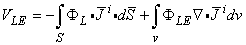
VLE = -F2 · 0(11.47) Note that -F2 corresponds precisely to a description of the sensitivity distribution associated with this particular lead, and is in fact the lead vector (field). Since no assumption has been made concerning the volume conductor, we have found a powerful method for quantitatively evaluating lead vector fields of arbitrary leads on arbitrary shaped inhomogeneous volume conductors.
The actual bioelectric sources may be characterized as a volume distribution i with dimensions of current dipole moment per unit volume. Equation 11.47 may be generalized to the case of such a volume distribution of current dipoles with a dipole moment density of i to obtain
(11.48) The quantity F2 was earlier defined as the reciprocal electric potential field in the volume conductor due to unit reciprocal current flow in the pickup leads a and b and is designated in the following as FLE. Plonsey (1963) has termed this potential field as the lead field in his field-theoretic proof of the reciprocity theorem. In this text, however, the term "lead field" denotes the current density field due to reciprocal application of current in the lead. They are related, of course, by LE = -sFLE.
Using the vector identity · (FLEi ) = FLE · i + FLE · i and the divergence theorem, we obtain from Equation 11.48
(11.49)
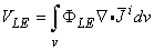
(11.50) · i is the strength of the impressed current source and is called the flow (or flux) source IF as defined in Equation 8.35. Thus Equation 11.50 can be expressed as
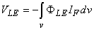
(11.51) McFee and Johnston (1953) designated the vector field LE = -sF = s LE the lead field. Here the symbol ELE denotes the reciprocal electric field due to unit reciprocal current, and the conductivity of the volume conductor. Using this formulation, we may rewrite Equation 11.48 as:
(11.52) LE is the lead field arising from unit reciprocal current (the reader should review the definition of J in Equation 11.41). But Equation 11.52 corresponds precisely to Equation 11.30 (assuming Ir = 1 [A]). Consequently, Equation 11.52 confirms Equation 11.30, which is the equation characterizing lead field theory, introduced earlier.
11.6.6 Summary of the Lead Field Theory Equations
In this section we summarize the equations of the lead field theory for electric leads. (Equations for magnetic leads are given in the next chapter.) We consider the situation in Figure 11.23, where two disklike electrodes in a volume conductor form the bipolar electric lead.
To determine the lead field, a unit reciprocal current Ir is fed to the lead. It generates a reciprocal electric potential field FLE in the volume conductor (this potential field was defined as F2 in Section 11.6.5 in the proof of the reciprocity theorem). If the electrodes are parallel and their lateral dimensions are large compared to their separation, FLE is uniform in the central region. The negative gradient of this electric potential field FLE is the reciprocal electric field, LE :
LE = – FLE (11.53) The reciprocal electric field is related to the reciprocal current field by the conductivity of the medium:
LE = sLE(11.54) Now, when we know the lead field LE, we can remove the reciprocal current generator (of unit current) from the lead. The electric signal VLE in the lead due to current sources i in the volume conductor is obtained from the equation
(11.30)
(11.31) Section 11.6.1 introduced the concept of isosensitivity surface and its special case half-sensitivity surface which bounds a half-sensitivity volume. The isosensitivity surfaces, including the half-sensitivity surface, are surfaces where the lead field current density LE is constant. In a homogeneous region of a volume conductor, where s is constant, the isosensitivity surfaces are, of course, surfaces where the reciprocal electric field LE is constant. In certain cases the isosensitivity surfaces coincide with the isopotential surfaces. These cases include those, where all isopotential surfaces are parallel planes, concentric cylinders, or concentric spheres. Then the surfaces where the electric field is constant (i.e. where two adjoining isopotential surfaces are separated by a constant distance) have the same form as well. But in a general case, where the isopotential surfaces are irregular so that two adjacent surfaces are not a constant distance apart the surfaces of constant electric field do not have the same form.
As summarized in Figure 11.23, as a consequence of the reciprocal energization of an electric lead, the following three fields are created in the volume conductor: electric potential field FLE (illustrated with isopotential surfaces), electric field LE (illustrated with field lines) and current field LE (illustrated with current flow lines and called the lead field). In addition to these three fields we defined a fourth field of surfaces (or lines): the field of isosensitivity surfaces. When the conductivity is isotropic, the electric field lines coincide with the current flow lines. In a symmetric case where all isopotential surfaces are parallel planes, concentric cylinders, or concentric spheres, the isopotential surfaces and the isosensitivity surfaces coincide.
In an ideal lead field for detecting the equivalent dipole moment of a volume source (see the following section) the isopotential surfaces are parallel planes. To achieve this situation, the volume conductor must also be homogeneous. Thus, in such a case from the aforementioned four fields, the electric field lines coincide with the lead field flow lines and the isopotential surfaces coincide with the isosensitivity surfaces..
Fig. 11.23. Basic form of a bipolar electric lead, where
| Ir | = unit reciprocal current; | |
| FLE | = reciprocal electric scalar potential field; | |
| LE | = reciprocal electric field; | |
| LE | = lead field: | |
| VLE | = voltage in the lead due to the volume source i in the volume conductor; and | |
| s | = conductivity of the medium. |
In this section we determine the desired form of the lead field of a detector that measures the equivalent (resultant) electric dipole moment of a distributed volume source located in an infinite homogeneous volume conductor.
As discussed in Section 7.3.2, a dipole in a fixed location has three independent variables, the magnitudes of the x-, y-, and z-components. These can be measured with either unipolar or bipolar electrodes locating at the coordinate axes. The vectorial sum of these measurements is the dipole moment of the dipole.
Because a volume source is formed from a distribution of dipole elements, it follows from the principle of superposition that the dipole moment of a volume source equals to the sum of the dipole moments of its dipole elements. This can be determined by measuring the x-, y-, and z-components of all the elementary dipoles and their sums are the x-, y-, and z-components of the equivalent dipole moment of the volume source, respectively. To introduce the important equations we show this fact also in mathematical form.
The equivalent electric dipole moment of a volume source may be evaluated from its flow source description. It was shown in Equation 8.35 (Section 8.5) that the flow source density IF is defined by the impressed current density (Plonsey, 1971) as
| IF = – · i | (8.35) |
The resultant (electric) dipole moment of such a source can be shown to be
 | (11.55) |
This dipole moment has three components. Because = x + y + z, these three components may be written as:
 | (11.56) |
We consider the x-component of the dipole moment. Noting Equation 8.35 and using the vector identity · (xi) = x · i + i · x, we obtain
 | (11.57) |
Using the divergence theorem, we may rewrite the first term on the right-hand side of Equation 11.57 as
 | (11.58) |
Since there can be no impressed current density on the surface, this term vanishes. Therefore, and because x = ( x/x) + (y/y) + (z/z) = i , we obtain for the x-component of the dipole moment
 | (11.59) |
In fact, recalling the dual identity of The lead field current density is given by The lead field current density is uniform throughout the source area.
Three such identical, mutually perpendicular lead fields form the three orthogonal components of a complete lead system.
i as a dipole moment per unit volume, we can write Equation 11.59 directly.
Equation 11.59 can be described as follows: one component of the equivalent electric dipole (moment) of a volume source may be evaluated from the sum of corresponding components of the distributed dipole elements of the volume source independent of their location. A comparison of Equation 11.59 with Equation 11.49 identifies FL with –x. Consequently, we see that this summation is, in fact, accomplished with a lead system with the following properties (see Figure 11.24):
L = x = (so that it is everywhere in the x direction only).
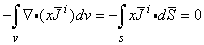
Fig. 11.24. Ideal lead field (sensitivity distribution) for detecting the electric dipole moment of a volume source. Each component is uniform in one direction throughout the source region, and the components are mutually orthogonal.
(A) Lead field current density vector presentation.
(B) Lead field current flow line presentation.
This is the physiological meaning of the measurement of the electric dipole. (See the text for details.)
The concept "physiological meaning" can be explained as follows: When considering the forward problem, the lead field illustrates what is the contribution (effect) of each active cell on the signals of the lead system. When one is considering the inverse problem, the lead field illustrates similarly the most probable distribution and orientation of active cells when a signal is detected in a lead..11.6.8 Application of Lead Field Theory to the Einthoven Limb Leads
PRECONDITIONS:
SOURCE: Volume source
CONDUCTOR: Infinite, homogeneous
To build a bridge between lead field and lead vector and to clarify the result of Equation 11.59 illustrated in Figure 11.24, we apply lead field theory to the Einthoven limb leads.
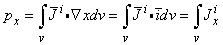
Previously, in Section 11.4.3, the Einthoven triangle was discussed as an application of the lead vector concept. The volume source of the heart was modeled with a (two-dimensional) dipole in the frontal plane. It was shown that the signal in each limb lead VI, VII, and VIII is proportional to the projections of the equivalent dipole on the corresponding lead vectors.
Instead of modeling the volume source of the heart with the resultant of its dipole elements, we could have determined the contribution of each dipole element to the limb leads and summed up these contributions. In this procedure one can use the lead field theory to illustrate the lead fields - that is, the sensitivities of the limb leads. The idealized lead fields of the limb leads are uniform in the directions of the edges of the Einthoven triangle. Figure 11.25 illustrates the sensitivity distribution of the (ideal) Einthoven limb leads within the area of the heart.
This is the physiological meaning of the measurement of the Einthoven limb leads (see the previous section).
Fig. 11.25. The ideal lead field (sensitivity distribution) of Einthoven limb leads VI, VII, and VIII. This is the physiological meaning of the measurement of the limb leads.
We begin the discussion on synthesis of ideal lead fields for detecting the equivalent dipole moment of a volume source by discussing the properties of unipolar and bipolar leads in infinite, homogeneous volume conductors.
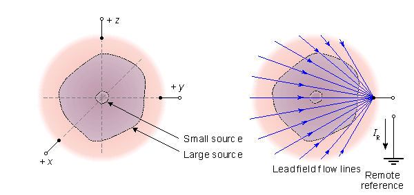
If the dimensions of a distributed volume source are small in relation to the distance to the point of observation, we can consider it to be a lumped (discrete) dipole. The detection of such an electric dipole is possible to accomplish through unipolar measurements on each coordinate axis, as illustrated on the left hand side of Figure 11.26A. If the dimensions of the distributed volume source are large in relation to the measurement distance, the lead field of a unipolar measurement is not directed in the desired direction in different areas of the volume source and the magnitude of the sensitivity is larger in the areas closer to the electrode than farther away. This is illustrated on the right hand side of Figure 11.26A.
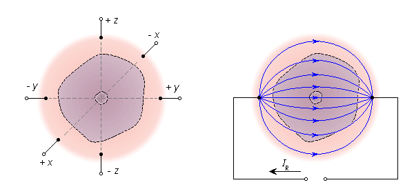
The quality of the lead field both in its direction and its magnitude is considerably improved when using a bipolar lead, where the electrodes are located symmetrically on both sides of the volume source, as illustrated in Figure 11.26B. (Note also that in the bipolar measurement the difference in potential between the electrodes is twice the unipolar potential relative to the center.)
The quality of the lead field of a bipolar lead in measuring volume sources with large dimensions is further increased by using large electrodes, whose dimensions are comparable to the source dimensions. This is illustrated in Figure 11.26C.
| ELECTRODE CONFIGURATION | LEAD FIELD OF ONE COMPONENT | ||
| A | UNIPOLAR LEADS, POINT ELECTRODES | ||
 | |||
| B | BIPOLAR LEADS, POINT ELECTRODES | ||
 | |||
| C | BIPOLAR LEADS, LARGE ELECTRODES | ||
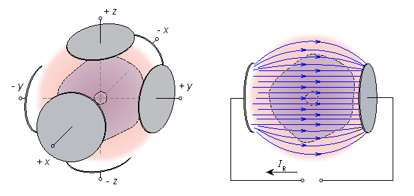 | |||
Fig. 11.26. Properties of unipolar and bipolar leads in detecting the equivalent electric dipole moment of a volume source.
(A) If the dimensions of the volume source are small compared to the measurement distance the simplest method is to use point electrodes and unipolar leads on the coordinate axes.
(B) For volume sources with large dimensions the quality of the lead field is considerably improved with the application of bipolar leads.
(C) Increasing the size of the electrodes further improves the quality of the leads.
Using large electrodes is in practice impossible. In the following we describe a method to design a lead to detect the equivalent electric dipole moment of a volume source in a finite, homogeneous volume conductor of arbitrary shape (Brody, 1957).
According to Section 11.6.7, such a lead, when energized reciprocally, produces three orthogonal, uniform, and homogeneous lead fields. We consider the construction of one of them. This may be done according to the following steps:
Suppose that the volume conductor has the arbitrary shape shown in Figure 11.27A and that our purpose is to synthesize an ideal lead field in the y direction within this region.
We extend the volume conductor in the direction of the y-axis in both directions so that it forms a cylinder limited by two planes in the zx direction and having the cross section of the original volume conductor (Figure 11.27B).
Then we plate the end planes of the cylinder with a well-conducting material. If electrodes are connected to these plates and a reciprocal current is fed to them, an ideal lead field is created in the volume conductor (Figure 11.27B).
Thereafter the extension of the volume conductor is slit as described graphically in Figure 11.27C, generating isolated "fibers." These cuts do not modify the form of the lead field because they are made along the flow lines which are nowhere intersected, as is clear in Figure 11.27C.
Each of the volume conductor "fibers" may now be replaced with discrete resistances of equal resistive value, as illustrated in Figure 11.27D.
The above procedure is repeated in the direction of the z- and x-axes. Corresponding to each discrete resistor, an electrode must be placed on the volume conductor. If the number of electrodes is sufficiently large, the ideal lead field (requiring an infinite number of electrodes) will be well approximated. Since one wishes to keep the number of electrodes to a minimum, one must explore the acceptability of reduced numbers of electrodes, make the spacing of electrodes unequal to strengthen accuracy only in the heart region, and use the same electrode for more than a single component lead.
Note once again, that this method may be applied to a finite homogeneous volume conductor having an arbitrary shape. In general, the effect of internal inhomogeneities cannot be corrected with electrodes located on the surface of the conductor with the method described above.
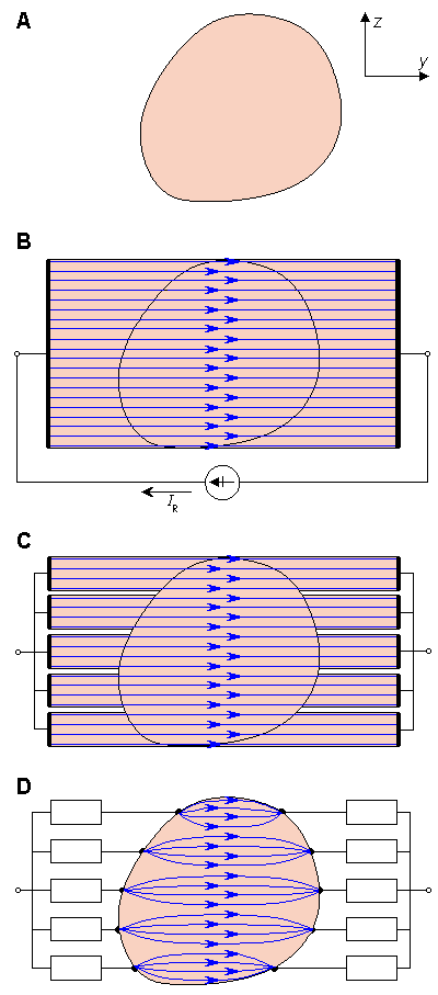
If the volume conductor is cut or an inhomogeneity boundary is inserted along a lead field current flow line, the form of the lead field does not change. Only the intensity of the field changes in relation to the conductivity.
The reciprocity theorem may be applied to the reciprocal situation. This means that it is possible in electrolytic tank models to feed a "reciprocally reciprocal" current to the dipole in the conductor and to measure the signal from the lead and interpret the result as having been obtained by feeding the reciprocal current to the lead and measuring the signal from the dipole.
The latter is easily proved by imagining that the lead field is a result of the mapping of the behavior of the lead vector as a function of the source location, as discussed in Section 11.6.2. This mapping is done by feeding unit currents in each coordinate direction at each point of the source area and by measuring the corresponding voltages at the lead, as explained in Section 11.4.1.
The benefit of this "reciprocally reciprocal" arrangement is that for technical reasons, the signal-to-noise ratio of the measurement may be improved while still having the advantage of the interpretations associated with the lead field current distribution.
The special properties of electric lead fields are discussed in more detail in connection with magnetic lead fields.
The source in the concept of the image surface is a dipole. This can be a discrete dipole (at a point), or it can be a dipole element of a distributed volume source. In the lead field concept, the source may be a distributed volume source or a discrete dipole. The conductor in both cases was previously considered to be finite (and inhomogeneous). However, the theory holds for infinite volume conductors as well.
Source location in the image surface was fixed and the measurement points were variable and forming a continuum. In characterizing the lead field, we note that the situation is the opposite: The measurement points are fixed while the source point varies (continuously). This means that in the image surface the lead vectors are mapped as a function of the measurement point, but in the lead field the mapping is a function of the source point. The image surface takes into account field points lying on a surface, whereas in the lead field the source point may lie within a three-dimensional volume.
Geometrically, in the image surface concept, the ends of the lead vectors form the image surface. In the lead field concept, the field of lead vectors establish the lead field.
The equations for the application of the lead vector and image surface (Equation 11.16) and the lead field (Equations 11.30 and 11.31) are, in principle, of the same form. The main difference is that the equation for the lead field is in integral form. This comes from the fact that it is applied to a volume source.
An important consequence of the reciprocity theorem of Helmholtz is that the lead field is identical to the current field resulting from feeding the (unit) reciprocal current to the lead.
| ||||||||||||||||||||||||||||||||||
| Note: | (1) There is similarity between the variables:  LE , i LE , i | |||||||||||||||||||||||||||||||||
| (2) It follows from the reciprocity theorem that i is the same as the current density field in the volume conductor due to feeding the reciprocal current Ir of 1 A to the lead. | ||||||||||||||||||||||||||||||||||
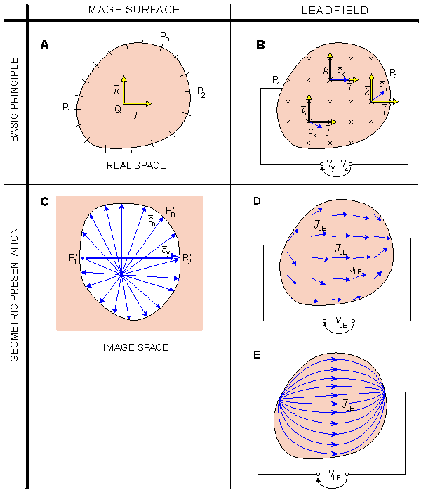
As described in Section 8.5 (Equation 8.35), the flow (flux) source density IF of a distribution of impressed current density i is
| IF = – · i | (8.35) |
and the resulting (electric) dipole moment of such a system is evaluated from the definition (Equation 11.55)
| (11.55) |
| where | | = the radius vector |
| dv | = the volume element |
The dipole moment has three components, as was illustrated by Equation 11.56. We now examine the x-component of this dipole moment. We develop it in the following way: The explanation for each step is given on the right-hand side of the column.
| px |  | from Equation 11.57 |
 | because –IF = · i (Equation 8.35) | |
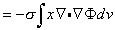 | from Equation 7.3 we have · i = · sF ; for a uniform conducting medium this reduces to · i = s · F | |
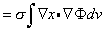 | · (xF) = x · F + x · F is a vector identity. Integrating each term through the entire volume, and applying Gauss's theorem to the first integral, we get 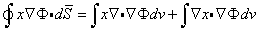 Since the boundary is insulated,F · d Since the boundary is insulated,F · d = 0 . Thus = 0 . Thus 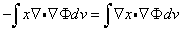 | |
 | 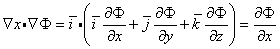 | |
 | because the volume integral may be transformed to a surface integral by integrating with respect to x | |
 | because the surface integral may be written in a more convenient form by using a vectorial surface element dx whose absolute value is dS = dy dz, and which is directed outward and normal to the surface defined by dy dz. |
which expresses the resultant dipole moment of a volume source in an arbitrary volume conductor.
Summing Equation 11.61 and similar expressions for py and pz and replacing the potential F with voltage V, we finally obtain the vector equation

(11.62) We now explain in detail the meaning of Equation 11.62, as illustrated in Figure 11.29.
Figure 11.29A illustrates the homogeneous volume conductor including the volume source. In the illustration the Gabor-Nelson theorem is discussed in two dimensions. The equivalent dipole moment of the volume source is . The vectorial surface element d is a vector attached to the surface element. It is directed outward and normal to the surface element, and its absolute value equals the area of the surface element. For clarity the volume conductor is divided into 12 surface elements, S1 through S12. (When applying Equation 11.62, of course, one assumes that the number of surface elements is infinite.) A vector d1 through d12 is attached to each surface element. The volume source produces a potential, F1 through F12, at each surface element.
It is obvious that because the surface is closed, the sum of the vectorial surface elements is equal to zero; that is, Sdi = 0 (Figure 11.29B).
If we multiply each vectorial surface element di by the corresponding potential Fi (or actually with the voltage Vi measured at each surface element in relation to an indifferent reference), the sum of these products, SVidi, is no longer zero. It is clear that when one is considering the surface potential due to the dipole along the surface elements of increasing index, one finds that it is at its maximum at the surface elements S1 and S2. Then it decreases and reaches the value zero somewhere between the surface elements S4 and S5. Thereafter the surface potential turns to negative polarity and reaches its maximum at the surface element S7. Thereafter the (negative) surface potential decreases to zero and increases again to the positive maximum in the area of S1. Therefore, the sum SVidi is not zero; and according to the Equation 11.62, if the number of the surface elements is infinite, one obtains . For clarity, the length of is shown longer in Figure 11.29C than in Figure 11.29A..
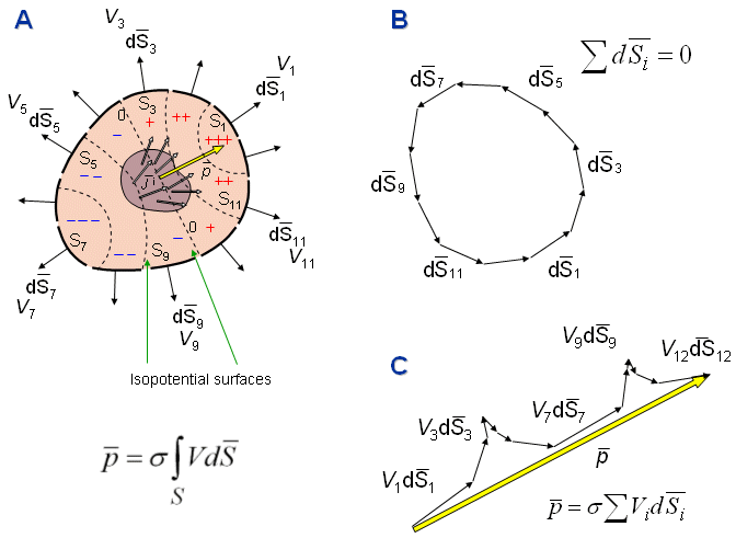
Fig. 11.29. Illustration of the Gabor-Nelson equation for evaluating the resultant (equivalent) dipole of a volume distribution lying in a bounded homogeneous volume conductor. The integral is shown approximated by discretizing the surface into 12 elements. The calculation of the integral is explained in detail in the text.
 |
the second moment of the source distribution is in the x direction and given by
 | (11.63) |
where upper-case X denotes the x-coordinate of the dipole location and lower-case x is the variable in this coordinate.
Integrating by parts with respect to x and again replacing the potential F with the measured voltage V gives
Two similar equations arise by cyclic permutation of the coordinates. We can also derive three new equations of the type
in the same manner as Equations 11.61 and 11.66 were derived. We can now eliminate the unknown volume integral of V from the equations of the type 11.66, and, together with the three equations of the type 11.68, we are left with the five equations for the three quantities X, Y, and Z. Any three of these five equations can be used for finding the location of the dipole, and the other two for checking how well the assumption of one dipole accounts for the observation. One can also use the method of least squares to obtain the best fit.
1. For the forward problem:
2. For the inverse problem:
In various cases we had the following sources:
These sources have been located in volume conductors that were:
Fig. 11.30. (A) The source-conductor combinations where the solid angle theorem and Miller-Geselowitz model may be applied in solving the forward problem. (B) The source-conductor combinations where the lead vector, image surface, and lead field methods as well as Gabor-Nelson theorem may be applied in solving the inverse problem.
Brody DA (1957): A method for applying approximately ideal lead connections to homogeneous volume conductors of irregular shape. Am. Heart J. 53:(2) 174-82.
Burger HC (1967): Heart and Vector, (Series ed, HW Julius Jr.: Philips Technical Library.) 143 pp. Gordon & Breach Science Publishers, New York.
Burger HC, van Milaan JB (1946): Heart vector and leads - I. Br. Heart J. 8:(3) 157-61.
Burger HC, van Milaan JB (1947): Heart vector and leads - II. Br. Heart J. 9: 154-60.
Burger HC, van Milaan JB (1948): Heart vector and leads - III. Br. Heart J. 10: 233.
Einthoven W (1908): Weiteres über das Elektrokardiogram. Pflüger Arch. ges. Physiol. 122: 517-48.
Frank E (1954): The image surface of a homogeneous torso. Am. Heart J. 47: 757-68.
Gabor D, Nelson CV (1954): Determination of the resultant dipole of the heart from measurements on the body surface. J. Appl. Phys. 25:(4) 413-6.
Gulrajani RM, Mailloux GE (1983): A simulation study of the effects of torso inhomogeneities on electrocardiographic potentials using realistic heart and torso models. Circ. Res. 52: 45-56.
Helmholtz HLF (1853): Ueber einige Gesetze der Vertheilung elektrischer Ströme in körperlichen Leitern mit Anwendung auf die thierisch-elektrischen Versuche. Ann. Physik und Chemie 89: 211-33, 354-77.
Horácek BM (1971): The effect on electrocardiographic lead vectors of conductivity inhomogeneities in the human torso. Dalhousie University, Halifax, Nova Scotia, pp. 182. (Ph.D. thesis)
Hyttinen JA, Eskola HJ, Sievänen H, Malmivuo JA (1988): Atlas of the sensitivity distribution of the common ECG-lead systems. Tampere Univ. Techn., Inst. Biomed. Eng., Reports 2:(2) 25+67.
Malmivuo JA (1976): On the detection of the magnetic heart vector - An application of the reciprocity theorem. Helsinki Univ. Tech., Acta Polytechn. Scand., El. Eng. Series. Vol. 39., pp. 112. (Dr. tech. thesis)
McFee R, Johnston FD (1953): Electrocardiographic leads I. Introduction. Circulation 8:(10) 554-68.
McFee R, Johnston FD (1954a): Electrocardiographic leads II. Analysis. Circulation 9:(2) 255-66.
McFee R, Johnston FD (1954b): Electrocardiographic leads III. Synthesis. Circulation 9:(6) 868-80.
Miller WT, Geselowitz DB (1978): Simulation studies of the electrocardiogram, I. The normal heart. Circ. Res. 43:(2) 301-15.
Plonsey R (1963): Reciprocity applied to volume conductors and the EEG. IEEE Trans. Biomed. Electron. BME-10:(1) 9-12.
Plonsey R (1971): The biophysical basis for electrocardiology. CRC Crit. Rev. Bioeng. 1: 1-48.
Stratton JA (1941): Electromagnetic Theory, McGraw-Hill, New York.
Waller AD (1889): On the electromotive changes connected with the beat of the mammalian heart, and on the human heart in particular. Phil. Trans. R. Soc. (Lond.) 180: 169-94.
Wikswo JP, Malmivuo JA, Barry WM, Leifer M, Fairbank WM (1979): The theory and application of magnetocardiography. In Advances in Cardiovascular Physics, Vol. 2, ed. DN Ghista, pp. 1-67, S. Karger, Basel.
Wilson FN, Macleod AG, Barker PS (1931): Potential variations produced by the heart beat at the apices of Einthoven's triangle. Am. Heart J. 7: 207-11.
In the limit Dx 0 and IDx px we obtain
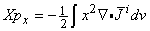
(11.64) We now transform this second moment integral, following the same steps as with the first moment, namely
Xpx
(11.65)
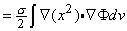

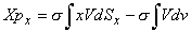
(11.66) In a similar way, we obtain equations for Ypy and Zpz. It is obvious that we cannot determine any of these by surface measurements alone because the second term in each expression requires a volume integral of the potential V. However, in the same way as one obtains Equation 11.66, one can show that
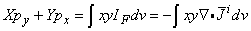
(11.67)
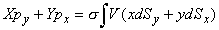
(11.68) 11.8 SUMMARY OF THE THEORETICAL METHODS FOR ANALYZING VOLUME SOURCES AND VOLUME CONDUCTORS
We have discussed six different theoretical methods for analyzing volume sources and volume conductors. Two of them are used for solving the forward problem, and the other four for solving the inverse problem. These methods are:
Solid angle theorem
Miller-Geselowitz model
Lead vector
Image surface
Lead field
Gabor-Nelson theorem
Double layer
Distributed dipole
Dipole (in a fixed location)
Moving dipole
Dipole moment of a volume source
Multiple dipole
Multipole
Infinite, homogeneous
(Infinite, inhomogeneous, not discussed)
Finite, homogeneous
Finite, inhomogeneous
The application of each method is limited to certain source-conductor combinations, as expressed in the sets of preconditions in connection with the discussion of each method. We summarize these preconditions in Figure 11.30. The former one of these shows the application areas for the two methods used in solving the forward problem, and the latter one for those used in solving the inverse problem.
Figure 11.30A is quite obvious. The preconditions of the two methods are shown by locating the methods in the corresponding location in the source-conductor plane. The application area of the solid angle theorem is shown to be both the infinite homogeneous volume conductor, as derived first by Helmholtz, and the finite homogeneous and inhomogeneous conductors, where it can be extended with the concept of secondary sources.
Figure 11.30B needs some clarification, and certainly some details of this figure could perhaps be presented also in some other way.
The source for the lead vector and image surface methods is dipole. In Sections 11.4 and 11.5, these methods were discussed, for simplicity, in connection with finite conductors. There is, however, no theoretical reason, that would restrict their application only to finite conductors, but they are applicable to infinite conductors as well. Therefore, their application area is shown for both finite and infinite conductors, but more light shaded in infinite conductors.
The same holds also for the lead field theory. Section 11.6.4 did not discuss the application of the lead field theory for a multiple dipole or multipole source. The lead field theory may, however, be applied also in connection with these sources. Therefore, they are included into the application area but with lighter shading.
The application area of the Gabor-Nelson theorem is clear. It can be applied for solving the dipole moment of a single dipole or a volume source in a finite homogeneous volume conductor. It also gives the location of this dipole moment..
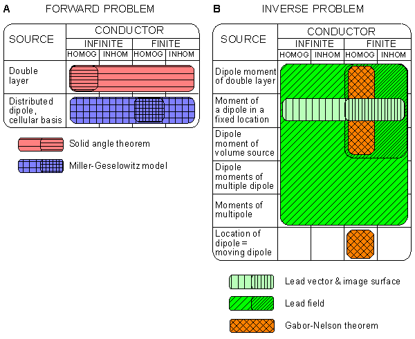
REFERENCES